IMAGE

Fig. 4

ID
ZDB-IMAGE-240621-82
Source
Figures for Wang et al., 2024
Image
Figure Caption

Fig. 4

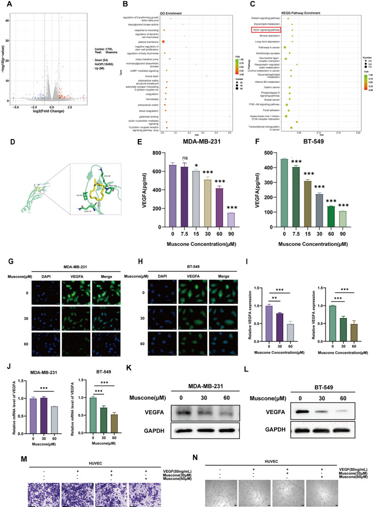
Muscone inhibits the VEGF axis in breast cancer (BC) cells. A Volcano plot of DEGs between control and muscone-treated MDA-MB-231 cells. Threshold was set at |log2FoldChange|> 1 and P < 0.05. Blue and red dots reflect diminished and elevated DEGs, respectively, by muscone treatment. B DEGs GO axis enrichment assessment. C DEGs KEGG network enrichment assessment. D Muscone interaction modes and locations with VEGFA, as estimated by AutoDock Vina software. E, F VEGFA secretion from BC cells was quantified by ELISA. G, H Cellular immunofluorescence (IF) assessments were conducted to assess VEGFA protein content in muscone-treated BC cells; Scale bar, 20 µm. I, J Western blot assessment of the muscone-mediated regulation of VEGFA protein levels in BC cells. K, L VEGFA transcript expression in BC cells as detected by RT-qPCR. M Muscone-mediated regulation of VEGF165-stimulated HUVECs migration as detected via Transwell cell migration assay; Scale bar, 1250 µm. N The muscone-mediated regulation of VEGF165-induced tube formation of HUVECs; Scale bar, 100 µm. Data shown as mean ± SEM. n = 3. *P < 0.05, **P < 0.01, and ***P < 0.001

Acknowledgments
This image is the copyrighted work of the attributed author or publisher, and ZFIN has permission only to display this image to its users. Additional permissions should be obtained from the applicable author or publisher of the image. Full text @ Cancer Cell Int.