IMAGE

Fig. 3

ID
ZDB-IMAGE-240621-81
Source
Figures for Wang et al., 2024
Image
Figure Caption

Fig. 3

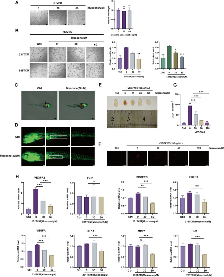
Muscone abrogates tumor angiogenesis in cellular and animal models. A, B Tube formation in vitro of HUVECs treated with muscone (A), or TCM from muscone-treated MDA-MB-231 cells (231TCM) or BT-549 cells (549TCM) (B). Tube lengths were measured with Fiji imaging software; Scale bar, 100 µm. C Equal numbers of Dil-labelled and incorporated MDA-MB-231 cells were administered into the zebrafish embryos perivitelline space at 48 h after fertilization. Arrows mark the administration locations. Magnification: 28.5 × . D Vasculature and neo-angiogenesis around the primary tumor and metastasis locations in zebrafish treated with muscone for 48 h. Boxes indicate newly formed tumor vessels. Magnification: 100 × . E Photographs of the Matrigel plugs from mice treated with VEGF and muscone for 12 d. F Representative images of immunofluorescent CD31+ microvessels (red) within Matrigel plugs; Scale bar, 100 µm. G Microvessel densities quantified as the density of CD31+ cells. H The effect of TCM from muscone-treated MDA-MB-231 cells (231TCM) on tumor angiogenesis-related gene expression in HUVECs. All data are shown as mean ± SEM, n = 3, *P < 0.05, **P < 0.01, and ***P < 0.001

Acknowledgments
This image is the copyrighted work of the attributed author or publisher, and ZFIN has permission only to display this image to its users. Additional permissions should be obtained from the applicable author or publisher of the image. Full text @ Cancer Cell Int.