
IMAGE
Fig. 1
- ID
- ZDB-IMAGE-240621-59
- Publication
- van Karnebeek et al., 2024 - CIAO1 and MMS19 deficiency: a lethal neurodegenerative phenotype caused by cytosolic Fe-S cluster protein assembly disorders
- All Figures
- Figures for van Karnebeek et al., 2024
Image
|
Figure Caption
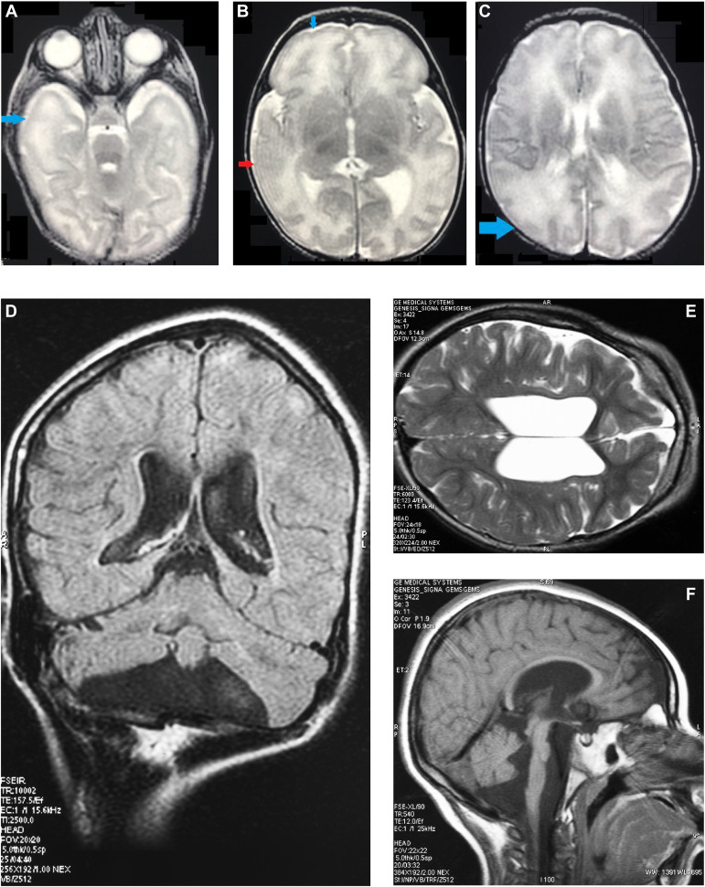
Fig. 1 Magnetic Resonance Images (MRI) of patient 1 and 3. T2-weighted images at the age of 5 days of patient 1 showed regions in both hemispheres with a simplified gyral pattern, indicated by blue arrows in (A), (B), and (C). The red arrow in (B) marks a possible abnormal lateral right temporal lobe, but the actual anatomy is obscured by a Gibbs artifact. MRI of patient 3 at the age of 4 years ([D] (FLAIR), [E] (T2 weighted), and [F] (T1 weighted) showed enlarged lateral and third ventricles, decreased volume of cerebral white matter and incomplete myelination, and pontocerebellar atrophy.
Acknowledgments
This image is the copyrighted work of the attributed author or publisher, and
ZFIN has permission only to display this image to its users.
Additional permissions should be obtained from the applicable author or publisher of the image.
Full text @ Genet. Med.