
Image
|
Figure Caption
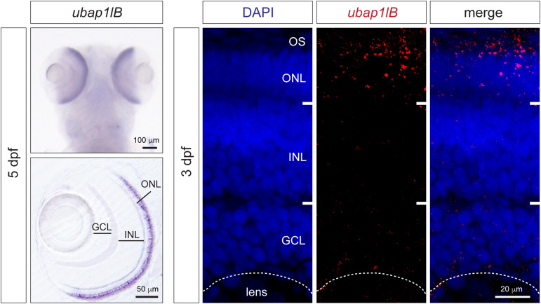
Fig. 4 Expression analysis of ubap1l in zebrafish. In situ hybridization for ubap1lb in zebrafish larvae 5 days after fertilization (left). The dorsal view is on the top and retina vibratome section is on the bottom. Expression can be detected in the retina in the photoreceptor layer. On the right, fluorescent HCR in situ hybridization on 3 days after fertilization larvae confirms ubap1lb mRNA expression in the OS and ONL where photoreceptors are located. ONL, outer nuclear layer; INL, inner nuclear layer; GCL, Ganglion cell layer; OS, outer segment.
Figure Data
Acknowledgments
This image is the copyrighted work of the attributed author or publisher, and
ZFIN has permission only to display this image to its users.
Additional permissions should be obtained from the applicable author or publisher of the image.
Full text @ Genet. Med.