
|
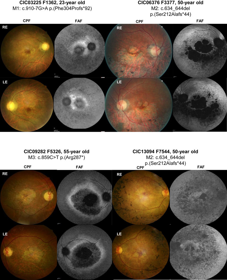
Fig. 2 Retinal images of index cases carrying homozygous variants in UBAP1L (CIC03225, CIC06376, CIC09282, and CIC13094). Color fundus photographs (CFP) and fundus autofluorescence imaging (FAF) of the right and left eye (RE and LE, respectively). CIC03225’s fundus examination revealed typical signs of RCD with pale optic discs, narrowed retinal vessels, and central macular atrophy and a loss of autofluorescence in the periphery, as well as in the macular region. CIC06376 showed central macular atrophy expending into the mid periphery predominantly in the inferior sector and a loss of autofluorescence in the atrophic areas surrounded by an area of heterogeneous autofluorescence expanding in the mid periphery. CIC09282 had pale optic discs, narrowed retinal vessels, and atrophic changes in the macular area with a loss of central autofluorescence surrounded by a ring of increased autofluorescence. CIC13094 had central macular atrophy and speckled appearance of autofluorescence at the posterior pole and mid periphery.