
|
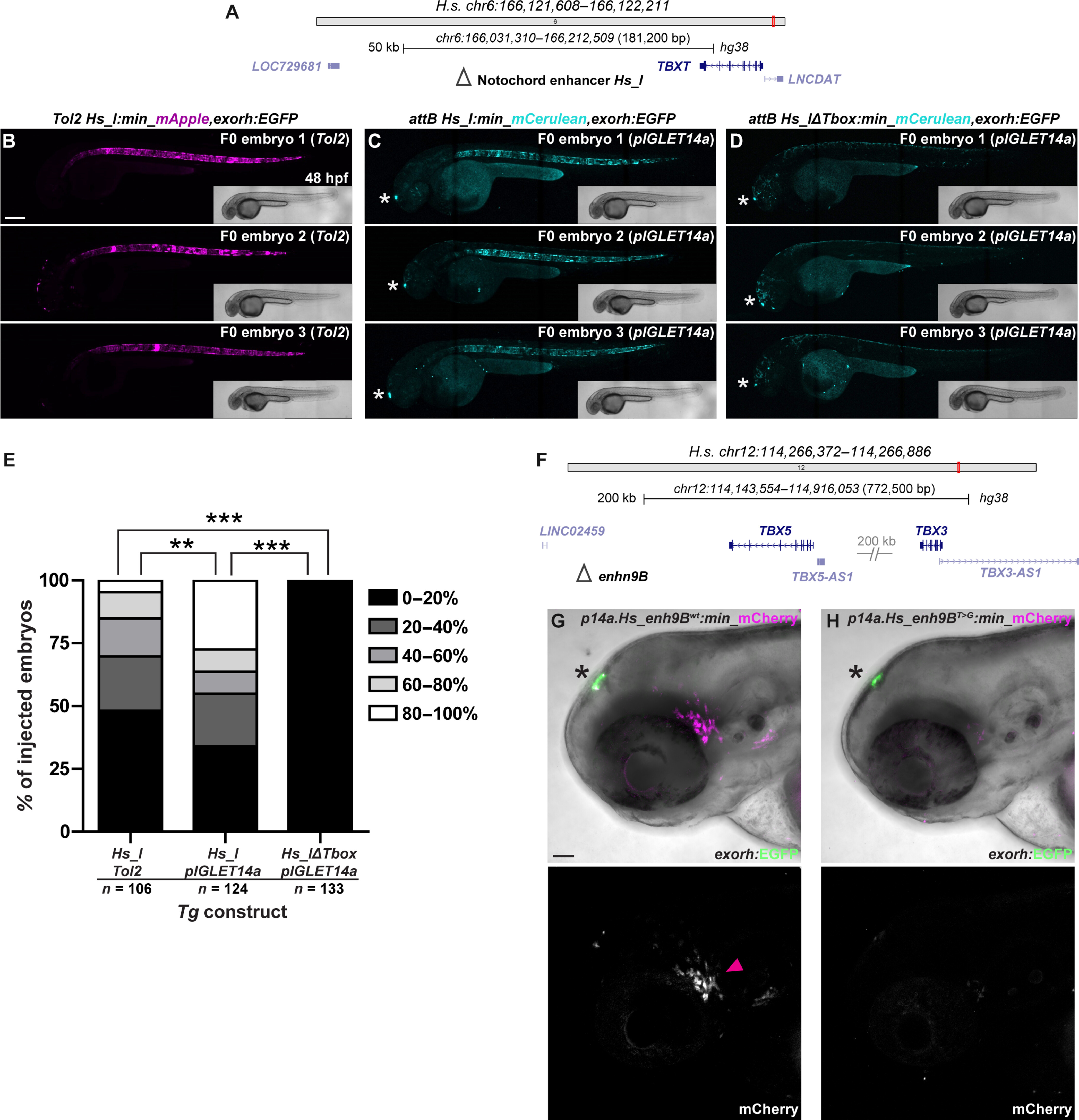
Fig. 4 pIGLET-based transgenesis facilitates reproducible enhancer variant testing in Zebrafish. (A to E) F0 injection–based testing of notochord enhancer Hs_I using Tol2- and pIGLET-based transgenesis. Schematic of TBXT locus with Hs_I annotated (A). Confocal imaging for mApple (B) or mCerulean (C and D) with bright-field inserts, anterior to the left. Both Tol2- and pIGLET14a-based F0 injections of Hs_I:min-mApple/mCerulean,exorh;EGFP result in embryos showing reporter expression with 80 to 100% fluorescent notochord coverage (B and C). Removal of the Tbox motif from Hs_I results in a complete loss of notochord reporter activity (D). Injected zebrafish are sorted for the exorh:EGFP transgenesis marker (white asterisk) to confirm successful injection (C and D). pIGLET14a-based F0 injections of Hs_I:min-mCerulean,exorh:EGFP show significantly more embryos with 80 to 100% fluorescent notochord coverage compared to Tol2-based injections and the Tbox motif deletion variant (E). (F to H) Comparison of stable reporter lines driven by the human TBX5/TBX3-associated enhancer 9B (Hs_enh9B, wt versus T > G variant). Schematic of TBX5/3 locus with enh9B annotated (F). Hs_enh9Bwt drives reporter expression in the ventral lateral line ganglia (magenta arrowhead) (G). Compared to wt, the disease-associated variant Hs_enh9BT>G exhibits nearly a complete loss of mCherry reporter expression (G and H). F1 heterozygous embryos are depicted (G and H). Scale bars: 200 μm (B) and 50 μm (G).