
|
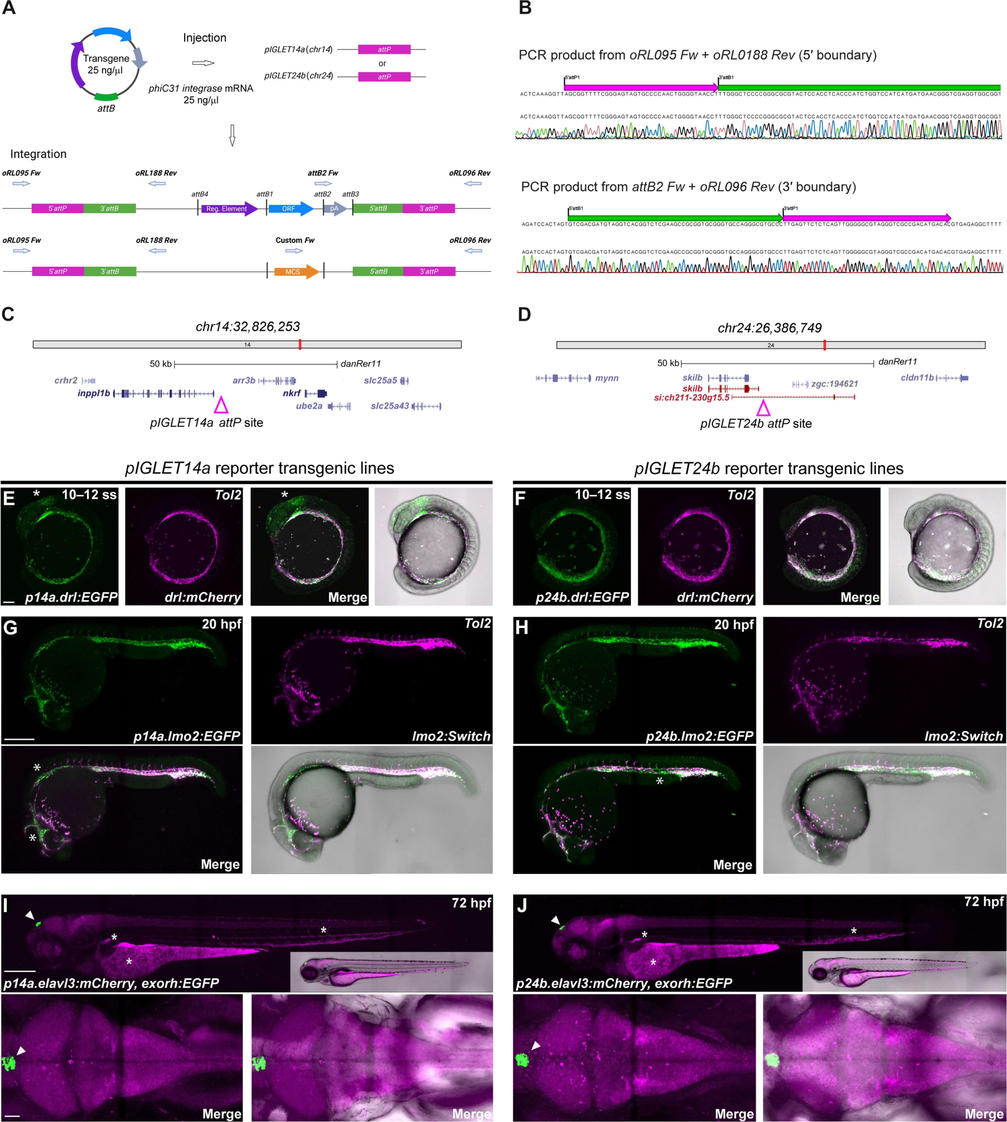
Fig. 1 Zebrafish pIGLET14a and pIGLET24b landing sites enable high-quality transgenics. (A and B) Schematic of pIGLET system workflow. Transgenes with attB-containing pDEST or MCS backbones are coinjected with phiC31 integrase mRNA into pIGLET14a or pIGLET24b zebrafish (A). Integration is depicted with both attB-containing pDEST (pCM268, pRL055, pRL056, pCK122, and pCK123) (top) or MCS (pRL092 and pRL093) (bottom) backbones (A). The 5′ and 3′ transgene integration boundaries can be confirmed via PCR and sequencing (B). (C and D) Genomic locations of both landing sites. (E to J) Validating pIGLET14a and pIGLET24b with F2 drl:EGFP, lmo2:EGFP,cryaa:Venus and elavl3:mCherry,exorh:EGFP. (E and F) Crossed to Tol2-based drl:mCherry, p14a.drl:EGFP and p24b.drl:EGFP show analogous reporter activity in lateral plate mesoderm (LPM; 10 to 12 ss green fluorescence). Note faint brain expression in p14a.drl:EGFP (E, white asterisk) common with Tol2-based drl reporter transgenics with strong expression. (G and H) Crossed to Tol2-based lmo2:Switch (dsRed2, magenta), p14a.lmo2:EGFP and p24b.lmo2:EGFP show analogous, overlapping activity in endothelium at 20 hpf. EGFP expression is more consistent/complete compared to dsRED2 in Tol2-based lmo2:Switch (G and H, white asterisk). (I and J) p14a.elavl3:mCherry and p24b.elavl3:mCherry show complete, consistent reporter expression in the central nervous system at 72 hpf, akin to previous Tol2-based elavl3 transgenics. The in cis exorh:EGFP transgenesis marker indicated with white arrowhead. Note common autofluorescence in blood, kidney, and yolk (I and J, white asterisk). F2 heterozygous embryos are depicted (E to J). Scale bars: 100 μm (E), 250 μm (G), 250 μm (I), and 50 μm.