
Fig. 6
- ID
- ZDB-IMAGE-240620-78
- Publication
- Du et al., 2024 - Lapatinib combined with doxorubicin causes dose-dependent cardiotoxicity partially through activating the p38MAPK signaling pathway in zebrafish embryos
- All Figures
- Figures for Du et al., 2024
|
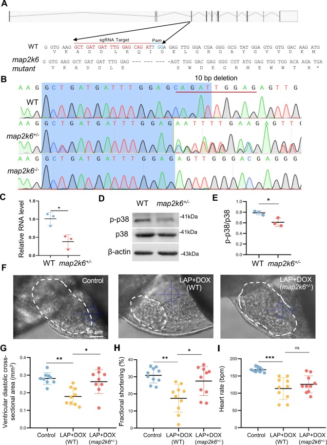
Fig. 6 The map2k6 frameshift mutation reduced combined LAP and DOX exposure-induced cardiotoxicity. (A) Schematic of the 10-nucleotide deletion allele generated by the injection of a single guide RNA (sgRNA) targeting the sequence within the 4th exon of the map2k6 gene. Dashed lines indicate deleted nucleotides. The asterisk indicates the predicted early translational stop codon. (B) Chromographs illustrating the sequences of WT and the map2k6 mutant allele with the 10-nucleotide deletion in F1 heterozygous and homozygous fish. (C) Quantitative RT-PCR demonstrated transcription reduction in the map2k6 heterozygous mutant. N=3 biological replicates. (D-E) Western blotting (D) and quantification (E) of p-p38 protein normalized by total p38 in map2k6 heterozygous mutant fish compared to that in WT Control hearts. N=3, Student’s t test. (F-I) Representative images (F) and quantification of the ventricular diastolic cross-sectional area (outlined by dashed white lines) (G), the percent fractional shortening (FS) (H) and heart rates (I) of the map2k6 heterozygous mutant embryos compared to WT embryos after exposure to LAP plus DOX or the DMSO control at 72 hpf. N=9–10, one-way ANOVA. *P<0.05, **P<0.01, ***P<0.001, ns, not significant; Scale bar: 50 µm.