
Fig. 1
- ID
- ZDB-IMAGE-240620-73
- Publication
- Du et al., 2024 - Lapatinib combined with doxorubicin causes dose-dependent cardiotoxicity partially through activating the p38MAPK signaling pathway in zebrafish embryos
- All Figures
- Figures for Du et al., 2024
|
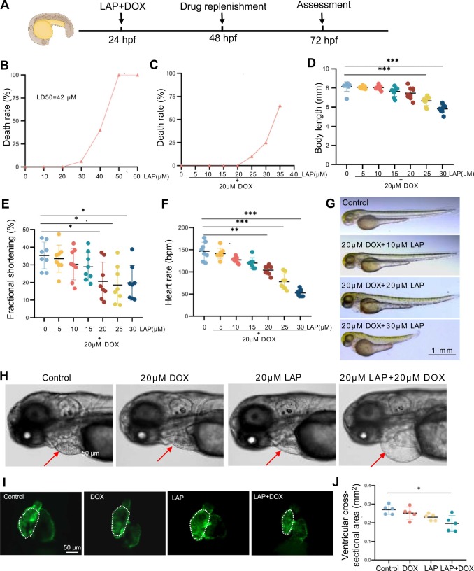
Fig. 1 Establishment of a dose-dependent cardiotoxicity model induced by lapatinib (LAP) combined with doxorubicin (DOX) in zebrafish embryos. (A) Experimental scheme showing combined LAP and DOX exposure to induce cardiotoxicity in zebrafish embryos. hpf: hours postfertilization. (B) Mortality of zebrafish embryos exposed to different doses of LAP alone. (C-D) Mortality (C) and body length (D) of zebrafish embryos exposed to different doses of LAP combined with 20 µM DOX. N=9, one-way analysis of variance (ANOVA). (E-F) Quantification of fractional shortening (FS) (E) and heart rate (F) in zebrafish embryos exposed to different doses of LAP combined with 20 µM DOX. bpm, beats per minute. N= 8, one-way ANOVA. (G-H) Representative images of embryonic morphology at 72 hpf after exposure to indicated doses of LAP and/or 20 µM DOX. The arrows point to the pericardium. (I-J) Representative fluorescence images of embryonic hearts (I) and quantification of the ventricular cross-sectional areas (J) of the Tg(cmlc2:EGFP) transgenic fish at 72 hpf after 20 µM LAP and 20 µM DOX combinational exposure compared to 20 µM LAP or 20 µM DOX alone or the DMSO control. N=5, one-way ANOVA; *P<0.05, **P<0.01, ***P<0.001.