
|
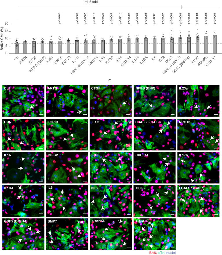
Fig. 2 Ability of candidate growth factors to induce neonatal cardiomyocyte cell-cycle progression Cardiomyocytes isolated from neonatal mice were cultured in vitro and stimulated for 48 h with selected growth factors (10 ng/mL), namely ARTN, CTGF, NPPB (BNP), IL23a, GDNF, FGF23, IL17f, LGALS3 (GAL3), NRG1b, IL1b, IGFBP, IL10, CXCL14, IL17b, IL1RA, IL6, IGF2, CCL3, GDF5 (BMP14), LGALS7 (GAL7), BMP7, sRANKL, and CXCL17. Cardiomyocytes were identified by cardiac Troponin I (cTnI) staining and analyzed by immunofluorescence for DNA synthesis (BrdU incorporation assay) (n = 34,914 cardiomyocytes pooled from the analysis of 162 samples); representative pictures are provided; arrows point at proliferating cardiomyocytes; scale bars, 20 μm. The values are presented as mean (error bars show SEM), statistical significance was determined using one-way ANOVA followed by Sidak’s test by comparing pairs of treatments (control vs. selected growth factor).