
|
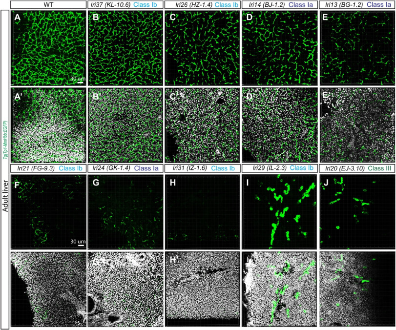
Fig. 7 Recovered viable mutants show distinguishable phenotypes in the adult liver. (A-J) Projected confocal images of the adult liver visualized for Tg(Tp1-MmHbb:EGFP)um14 expression in wild-type (A), lri37 (KL-10.6) (B), lri26 (HZ-1.4) (C), lri14 (BJ-1.2) (D), lri13 (BG-1.2) (E), lri21 (FG-9.3) (F), lri24 (GK-1.4) (G), lri31 (IZ-1.6) (H), lri29 (IL-2.3) (I), and lri20 (EJ-3.10) (J) mutant fish. DAPI staining is overlaid in (A′-J′). Cross sections of the liver were used. The histology of the liver based on DAPI is significantly altered in lri21 (F′), lri24 (G′) and lri21 (J′), possibly due to cholestasis. The z-thickness of all projected images is 0.42 μm.
Reprinted from Developmental Biology, 512, Singh, D.J., Tuscano, K.M., Ortega, A.L., Dimri, M., Tae, K., Lee, W., Muslim, M.A., Rivera Paz, I.M., Liu, J.L., Pierce, L.X., McClendon, A., Gibson, I., Livesay, J., Sakaguchi, T.F., Forward genetics combined with unsupervised classifications identified zebrafish mutants affecting biliary system formation, 44-56, Copyright (2024) with permission from Elsevier. Full text @ Dev. Biol.