
|
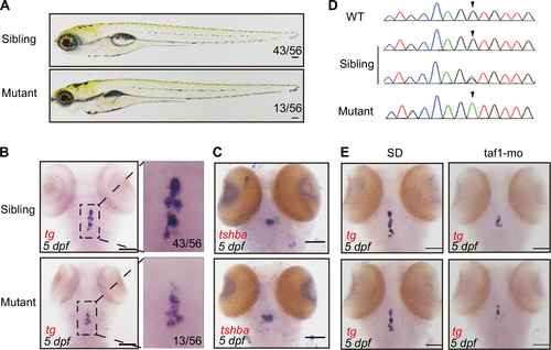
Fig. 1 Identification of taf1 mutations in thyroid mutants. A: the whole morphology of 031003 sibling vs. mutant larvae screened by N-ethyl-N-nitrosourea (ENU). B: thyroid marked by tg in 5 days postfertilization (dpf) embryos using whole mount RNA in situ hybridization (WISH). Nearly 25% of progeny from intercrossed heterozygotes showed an abnormal phenotype. Sibling contained wild-type and heterozygous embryos. C: the tshbα (thyrotrophin) in situ expression pattern in siblings and mutants. D: the 0310003 mutants had lesions in taf1 caused by V241M amino acid substitutions. Verification by Sanger sequencing. E: embryos injected with translation-blocking taf1 morpholinos (taf1-mo) can reproduce the small thyroid phenotype detected at 5 dpf by in situ hybridization. In the control group, embryos were injected with standard control morpholino (sd). Scale bar: 100 μm. tg, thyroglobulin.