IMAGE

Fig. 1

ID
ZDB-IMAGE-240614-47
Source
Figures for Miles et al., 2024
Image
Figure Caption

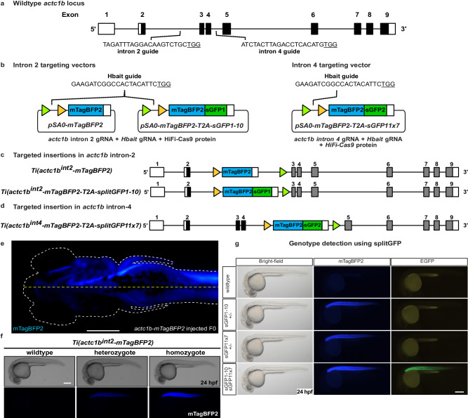
Fig. 1 SplitGFP alleles enable visual selection of homozygous mutants.

a Schematic of actc1b gene structure, and location of the intron 2 and intron 4 guideRNA sites. b Schematic of intron 2 and intron 4 targeting vectors, FRT and FRT3 sites are indicated by triangles. c Schematic of Ti(actc1bint2-mTagBFP2) and Ti(actc1bint2-mTagBFP2-T2A-sGFP1-10) intron 2 targeted insertion lines. d Intron 4 targeted Ti(actc1bint4-mTagBFP2-T2A-sGFP11x7) insertion line. e Example of successful targeted integration during the first cell division. Integration of the targeting vector into intron 2 of actc1b during the first cell division results in embryos with mTagBFP2 expression in one half of the ventral body plan. A white dotted line indicates embryo outline. The yellow dotted line indicates the ventral axis. In our experience, such embryos, when raised to adulthood and crossed, have always transmitted the successful integration event to their progeny. Ventral view of a 4 dpf embryo. f Brightfield and fluorescent images of wildtype, heterozygote, and homozygote embryos at 24 hpf. g 24 hpf embryos were generated by crossing the two actc1bsGFP lines together. Heterozygous carriers display mTagBFP2 expression only while, in addition to mTagBFP2 expression, compound heterozygous mutants can be visually selected by GFP fluorescence. Experiments were repeated three independent times. Scale bar = 250 μm.

Acknowledgments
This image is the copyrighted work of the attributed author or publisher, and ZFIN has permission only to display this image to its users. Additional permissions should be obtained from the applicable author or publisher of the image. Full text @ Nat. Commun.