IMAGE

Fig. 7

ID
ZDB-IMAGE-240612-26
Source
Figures for Akam-Baxter et al., 2024
Image
Figure Caption

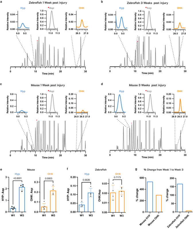
Fig. 7 Hydroxylation of lysine in newly deposited collagen differs markedly in infarcted murine and zebrafish hearts.

HPLC traces of the amino acids in infarcted zebrafish (a, b) and mouse (c, d) hearts at 1 and 3 weeks after injury. The expanded regions in the insets are for the hydroxyproline (Hyp) and hydroxylysine (OHK) peaks, at 9.3 min and 26.7 min respectively, and the Aspartic acid (Asp) peak at 11.3 min to which the chromatogram is normalized. Aspartic acid (Asp) was quantified and used to normalize Hyp and OHK values to account for total tissue content in each sample. The Asp peak is denoted with a red asterisks (*) in the full HPLC chromatograms. Quantification of Hyp:Asp and OHK:ASP in mouse (e, n = 5 for W1, n = 4 for W3) and zebrafish (f, n = 4) hearts at 1 and 3 weeks after injury. Hyp:Asp increases significantly in both zebrafish and mice, while OHK:Asp increases significantly in mice but not zebrafish. g Comparison of the percent change of Hyp and OHK from week 1 to week 3 after injury in mice and zebrafish hearts. Despite a marked increased in Hyp from weeks 1–3, indicative of new collagen deposition, little change in OHK is seen in the zebrafish hearts. Statistical comparison by unpaired t-test, n = 4–5 per sample values are plotted as the average ± the standard deviation. Source data are provided in the Source Data file.

Acknowledgments
This image is the copyrighted work of the attributed author or publisher, and ZFIN has permission only to display this image to its users. Additional permissions should be obtained from the applicable author or publisher of the image. Full text @ Nat. Commun.