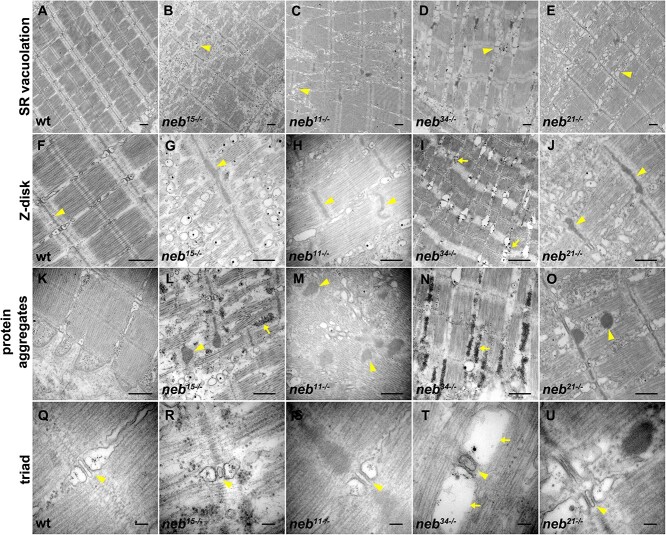
IMAGE

Figure 6

ID
ZDB-IMAGE-240607-122
Source
Figures for Fabian et al., 2024
Image
Figure Caption

Figure 6

Details of ultrastructural defects in neb mutants. (A–U) Transmission electron micrographs of zebrafish embryos at 6 dpf. (A) Aligned sarcomeres in wt muscle. (B–E) Increased vacuolation of the sarcoplasmic reticulum in neb15, neb11, neb34, neb21 mutants (arrowheads). (F) Z-disks in wt muscle (arrowheads). (G–J) Significantly thicker Z-disks in neb15, neb11, neb34, neb21 mutants (arrowheads). (K) No abnormal protein aggregates have been observed in wt muscle. (L, M, and O) Electron-dense structures with a fibrous composition were observed in neb11, neb15, neb21 (arrowheads). (L and N) Accumulation of ribosomes were observed in neb15 and neb34 mutants (arrows). (Q) Normal triad structure and organization in wt muscle (arrowhead). (R–U) Triad structure and organization did not seem to be defective in neb mutants (arrowheads), except in splice mutant neb34, where enlarged terminal cisternae were observed (arrows). Scale bars A-O = 500 nm. Scale bars Q-U = 100 nm.

Figure Data
Acknowledgments
This image is the copyrighted work of the attributed author or publisher, and ZFIN has permission only to display this image to its users. Additional permissions should be obtained from the applicable author or publisher of the image. Full text @ Hum. Mol. Genet.