IMAGE

Fig. 3.

ID
ZDB-IMAGE-240606-34
Source
Figures for Kaliya-Perumal et al., 2024
Image
Figure Caption

Fig. 3.

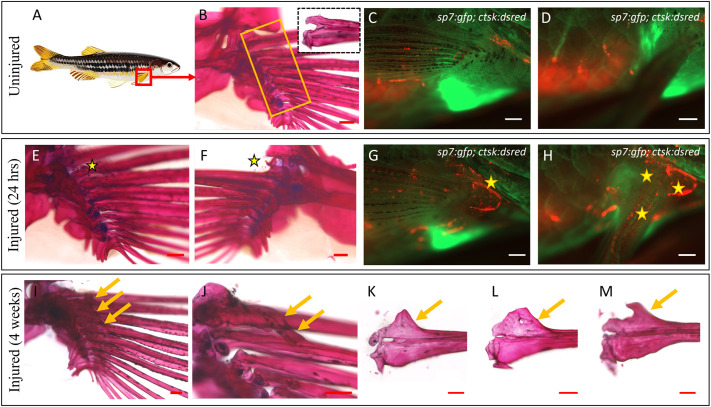
Consistent heterotopic ossification in the form of spurs and bridges in the pectoral fin. (A) Graphic depicting the injury site. (B) Medial view of an uninjured right pectoral fin stained with Alizarin Red. The yellow box indicates the injury site; the zoomed-in dotted box focuses on the articular region of a dissected fin ray, comprising the dorsal and ventral hemirays. (C) Lateral view of an uninjured pectoral fin of Tg(sp7:gfp; ctsk:dsred) fish under fluorescence microscopy. (D) The same fish as in C after elevating the pectoral fin to show the inner (medial) side where the absence of osteoclastic activity can be noted. (E,F) Medial and lateral views of an injured pectoral fin, 24 h post injury, showing tiny flecks of bone (stars) arising from the microfractures. (G) Lateral aspect of an injured pectoral fin of Tg(sp7:gfp; ctsk:dsred) fish showing osteoclastic activity (star) at 24 h post injury. (H) Osteoclastic activity on the medial aspect seen after elevating the fin (stars). (I) Injured pectoral fin, 1 month after injury. Arrows highlight the heterotopic bone spurs. (J) Heterotopic bridging bone noticed between the marginal ray and the second ray in one of the injured fish (arrows). (K-M) Different forms of heterotopic bone spurs encountered following injury (arrows). Images are representative of the first batch of five fish per condition. The observed heterotopic ossification phenotype was consistently reproducible in subsequent batches (n=32 fish). Scale bars: 200 µm (B,E,F,I-M); 500 µm (C,D,G,H).

Acknowledgments
This image is the copyrighted work of the attributed author or publisher, and ZFIN has permission only to display this image to its users. Additional permissions should be obtained from the applicable author or publisher of the image. Full text @ Dis. Model. Mech.