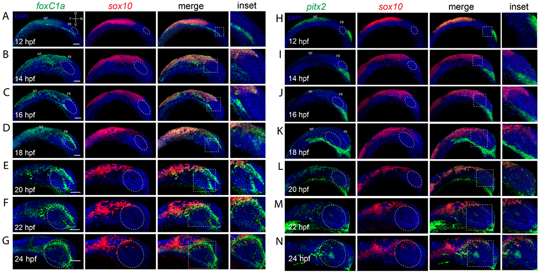
Fig. 3 Co-expression analysis of sox10 and anterior segment mesenchyme markers during eye field and retinal morphogenesis. A-G) Two color FWISH analysis of foxC1a (green) and sox10 (red) at 12-24hpf with samples taken at 2h intervals. DNA was stained with DAPI (blue). Lateral images of volume projections of 3D confocal stacks from representative embryos are displayed. Strong co-expression between sox10 and foxC1a is observed starting at 14hpf in the notochord. Anterior segment (white oval) expression for foxC1a is detected starting at 18hpf and continuing up to 24hpf while sox10 expression is excluded from the anterior segment region in all timepoints. H-N) Two color FWISH analysis of pitx2 (green) and sox10 (red) at 12-24hpf with samples taken at 2h intervals. DNA was stained with DAPI (blue). Lateral images of volume projections of 3D confocal stacks from representative embryos are displayed. Co-expression between sox10 and pitx2 is first observed starting at 20hpf in the notochord but overall co-expression is minor. Pitx2 expression is primarily constrained to regions ventral of the developing eye. By 22-24hpf pitx2 expression is detected in the anterior segment space (white oval) and includes several cells exhibiting co-expression with sox10. Scale bar = 100μm. NT = neural tube, FB = forebrain.
Image
Figure Caption
Acknowledgments
This image is the copyrighted work of the attributed author or publisher, and
ZFIN has permission only to display this image to its users.
Additional permissions should be obtained from the applicable author or publisher of the image.
Full text @ Differentiation

