
|
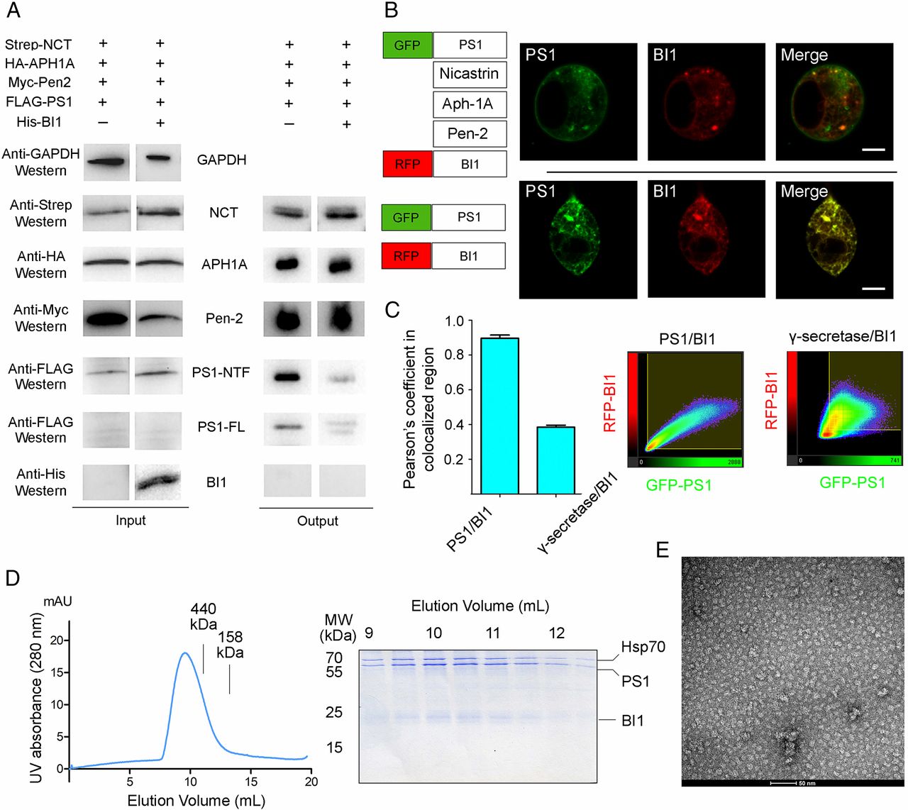
Fig. 4 BI1 binds the isolated PS1, but not PS1 in the context of an assembled γ-secretase. (A) BI1 is absent in the purified γ-secretase. (Left) All four components of γ-secretase and BI1 are readily detectable upon cellular overexpression. (Right) Following affinity purification of the FLAG-tagged PS1, the other three components of γ-secretase, but not BI1, are detectable by Western blot. (B) Colocalization of PS1 and BI1 is altered by expression of the other three components of. γ-secretase. (Top) Coexpression of GFP-PS1 and RFP-BI1 reveals excellent colocalization. (Bottom) Extent of colocalization is markedly reduced by coexpression of the other three components of γ-secretase (Scale bar, 10 μm.) (C) Quantification of colocalization between PS1 and BI1 by Pearson’s coefficient in colocalized volume. (Left) Coefficient in the presence of PS1 overexpression alone is considerably higher than that in the presence of overexpression by all four components of γ-secretase. (Right) Typical 2D correlation diagrams. (D) Purification of the PS1–BI1 complex for structural studies. (Left) Representative gel filtration chromatograph of the two-step purified human PS1–BI1 complex. (Right) Aliquots of the peak fractions are visualized in an SDS/PAGE gel. (E) Analysis of the purified PS1–BI1 complex by negative-staining EM. Shown here is a representative micrograph of the PS1–BI1 complex.