
|
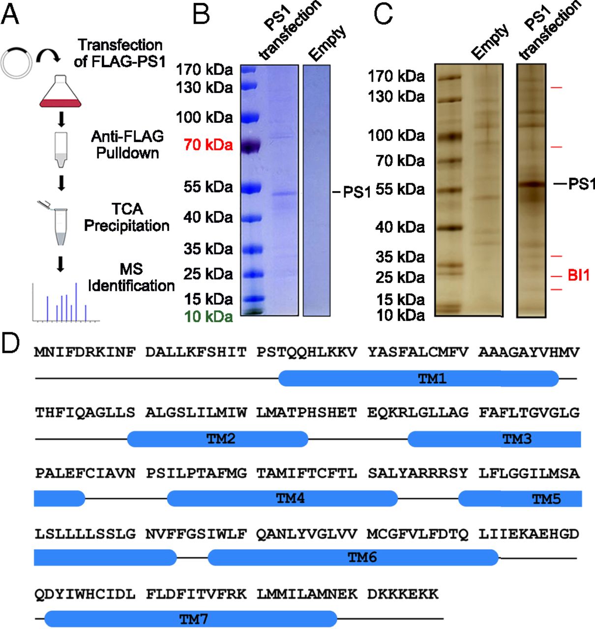
Fig. 1 Identification of BI1 as a potential PS1-binding protein. (A) Procedure for the identification of potential PS1-binding proteins. The FLAG-tagged human PS1 was overexpressed in HEK293F cells and purified by the M2 affinity resin. The eluted proteins were precipitated by trichloroacetic acid (TCA) and analyzed by MS. (B) Affinity-purified proteins were examined on an SDS/PAGE gel and stained by Coomassie blue. The full-length human PS1 is clearly present in the sample derived from PS1 transfection, but not in the sample derived from the empty vector control. (C) Affinity-purified proteins were examined by silver staining. A number of proteins are uniquely present in the sample derived from PS1 transfection. (D) Amino acid sequence of BI1. Blue cylinders denote predicted transmembrane helices (TMs) based on the structure of the bacterial BI1 homolog BsYetJ (60).