IMAGE

Fig. 3

ID
ZDB-IMAGE-240601-4
Source
Figures for Xiao et al., 2024
Image
Figure Caption

Fig. 3

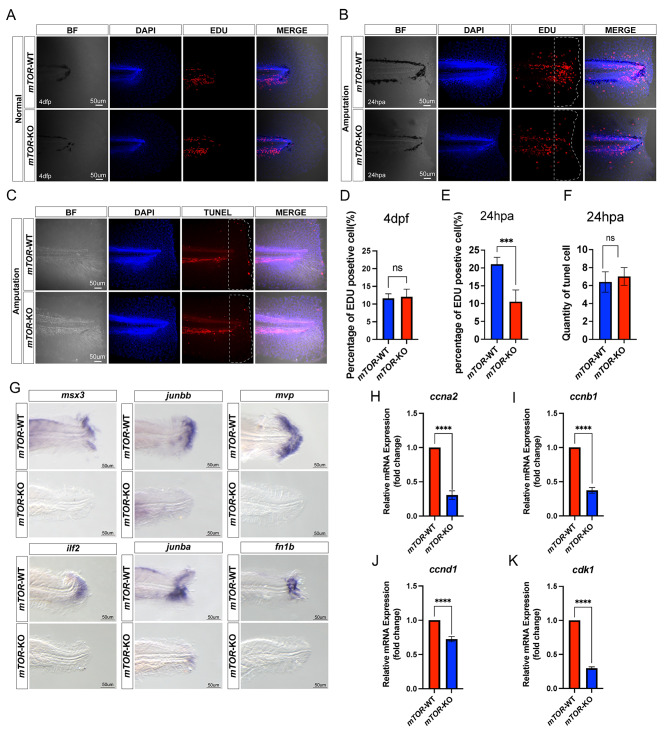
mTOR promoted epithelial and mesenchymal cells proliferation during larval zebrafish fin regeneration. (A, D) EDU staining of mTOR-WT and mTOR-KO larval zebrafish fin at 4 dpf. (B, E) EDU staining of mTOR-WT and mTOR-KO larval zebrafish fin at 24 hpa. (C, F) TUNEL staining of mTOR-WT and mTOR-KO larval zebrafish fin at 24hpa. (G) Location of msx3, junbb, mvp, ilf2, junba, fn1b in mTOR-WT and mTOR-KO larval zebrafish fin by in situ hybridization at 48 hpa. (H-K) The mRNA expression levels of cell cycle-related molecules (ccna2, ccnb1, ccnd1 and cdk1) between mTOR-WT and mTOR-KO larval zebrafish fin at 48 hpa. nsP > 0.05, ***P < 0.001, ****P < 0.0001

Acknowledgments
This image is the copyrighted work of the attributed author or publisher, and ZFIN has permission only to display this image to its users. Additional permissions should be obtained from the applicable author or publisher of the image. Full text @ J Orthop Surg Res