Image
Figure Caption

Figure 6—figure supplement 1. Examples of junctional contacts targeted by FRAP in the aortic landscape.

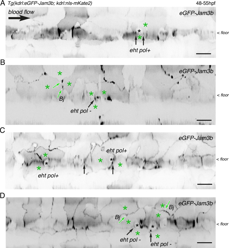
After performing z-stack acquisitions in trunk regions followed by 2D-deployment of aortic segments (A-C), 4 different Tg(kdrl:eGFP-Jam3b; kdrl:nls-mKate2) 48–55 hpf embryos were illuminated for FRAP. Panels (A and B) are the entire segments from which cropped images of EHT pol+ and EHT pol- cells (black asterisks) and surroundings were extracted to build the panels A and B of Figure 6. Panels (C and D) are from two other embryos. The 2D-cartographies allow visualizing the junctions that were selected for FRAP, with the black arrows pointing at junctional interfaces between EHT and endothelial cells (all are in the area of tri-junctions) and the green arrows pointing at bi-junctions (Bj) or tri-junctions between endothelial cells (green asterisks). Scale bars = 20 µm.

Acknowledgments
This image is the copyrighted work of the attributed author or publisher, and ZFIN has permission only to display this image to its users. Additional permissions should be obtained from the applicable author or publisher of the image. Full text @ Elife