|
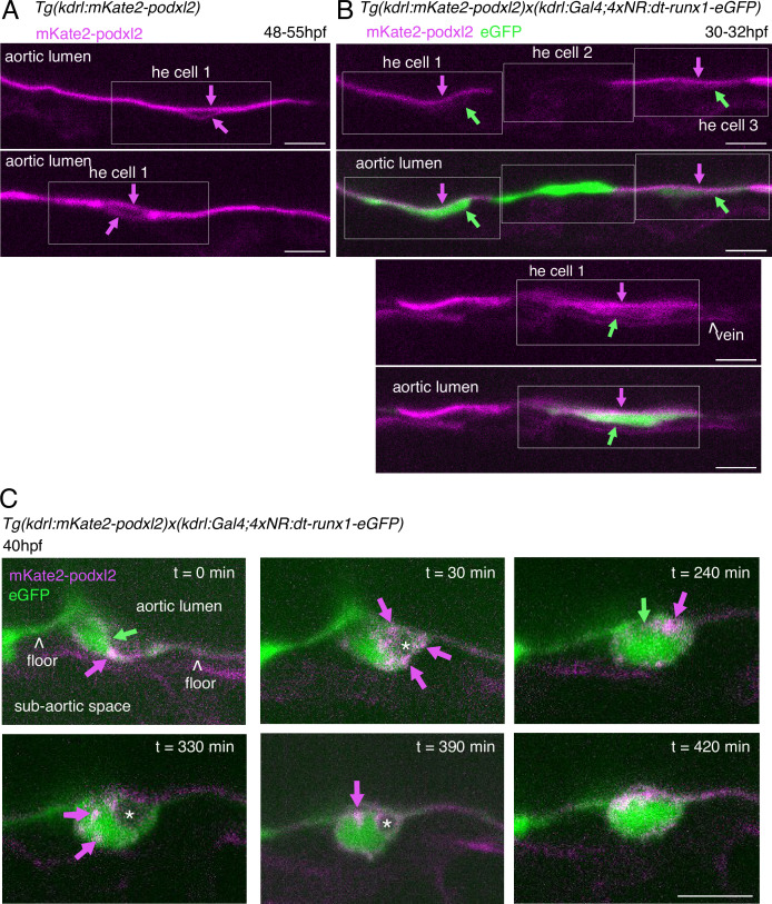
Figure 3—figure supplement 2. Phenotypic analysis of dt-runx1 expressing mutants: evidence for apicobasal polarity of hemogenic cells.
30–32 hpf and 48 - 55hpf embryos obtained from

|
|
Figure 3—figure supplement 2. Phenotypic analysis of dt-runx1 expressing mutants: evidence for apicobasal polarity of hemogenic cells.
30–32 hpf and 48 - 55hpf embryos obtained from