IMAGE

Fig 2

ID
ZDB-IMAGE-240524-80
Source
Figures for Li et al., 2024
Image
Figure Caption

Fig 2 Zebrafish mylipb exhibits proviral effects both in vivo and in vitro.

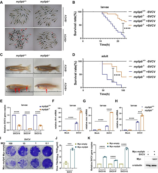
(A) Representative images of mylipb-null zebrafish larvae (mylipb-/-) (3 dpf) and WT siblings (mylipb+/+) (3 dpf) infected with or without SVCV for 18 h. Zebrafish larvae (3 dpf) were pooled in a disposable 60-mm cell culture dish filled with 5mL of egg water plus 2 mL SVCV (∼2.5×10⁷TCID50/ml). Dead larvae were identified by lack of movement, absence of blood circulation, and bodily degeneration (indicated by red arrows). (B) mylipb-null zebrafish (mylipb-/-) (n = 48, 3 dpf) were more resistance to SVCV infection than the WT (mylipb+/+) (n = 48, 3 dpf) based on the survival ratio. SVCV viruses (∼2.50 × 10⁷ TCID50/mL) were added to mylipb-null and WT larvae, and the numbers of dead larvae were counted every hour from 0 to 24 h. (C) mylipb-null zebrafish adults are more resistance to SVCV infection than WT zebrafish. mylipb-null zebrafish (mylipb-/-)(3 months postfertilization [3 mpf]) displayed less symptoms of abdominal hemorrhage after SVCV infection than WT zebrafish (mylipb+/+)(3 mpf). The zebrafish were i.p. injected with either 10 μL cell culture medium or 10 μL SVCV(∼2.50 × 10⁷ TCID50/mL). The red arrows indicate hemorrhage regions. (D) mylipb-null zebrafish (mylipb-/-) (3 mpf) (n = 10) were more resistance to SVCV infection than the WT (mylipb+/+) (3 mpf) (n = 10) based on the survival ratio. (E) The virus replication number was lower in mylipb-null zebrafish larvae than in the WT zebrafish after infection with SVCV. mylipb-null larvae (mylipb-/-) and the WT larvae (mylipb+/+) are offspring of siblings. 30 larvae (3 dpf) were pooled in a disposable 60-mm cell culture dish filled with 5 mL of egg water and 0.5 mL of SVCV (∼2.5×10⁷TCID50/ml).. After challenge for 24 h, the mRNA expression of N protein, P protein, and G protein of SVCV was detected by qRT-PCR analysis. (F-H) The induction of key antiviral genes, including mxc (F), ifn1 (G), lta (H) upon SVCV infection was higher in mylipb-null larvae (mylipb-/-) compared with the WT larvae (mylipb+/+). (I) Overexpression of mylipb reduced cell survival after SVCV infection in EPC cells. EPC cells were transfected with Myc-mylipb or empty vector. At 24 h post-transfection, the cells were infected with SVCV at the dose indicated for 48 h. Then, the cells were fixed with 4% paraformaldehyde and stained with 1% crystal violet. (J) Overexpression of mylipb increased virus titer in SVCV-infected EPC cells. The culture supernatant was collected from EPC cells infected with SVCV (MOI of 1), and the viral titer was measured by plaque assay. The results are representative of three independent experiments. (K) Overexpression of mylipb increased copy number of SVCV-related genes in SVCV-infected EPC cells. EPC cells were transfected with Myc-mylipb or empty vector for 24 h and infected with SVCV (MOI of 1). After 24 h, total RNAs were extracted for examining the mRNA levels of the N, P, and G gene of SVCV by qRT-PCR analysis. Western blotting tests to detect the overexpression of mylipb. All data are presented as mean values based on three repeated experiments, and error bars indicate the ± SD. *,P < 0.05, **, P<0.01; ***, P < 0.001; ****, P< 0.0001.

Acknowledgments
This image is the copyrighted work of the attributed author or publisher, and ZFIN has permission only to display this image to its users. Additional permissions should be obtained from the applicable author or publisher of the image. Full text @ PLoS Pathog.