
Fig. 5
- ID
- ZDB-IMAGE-240524-64
- Publication
- Fiegl et al., 2024 - Laboratory Course Using Zebrafish to Uncover Changing Roles of Wnt Signaling in Early Vertebrate Development
- All Figures
- Figures for Fiegl et al., 2024
|
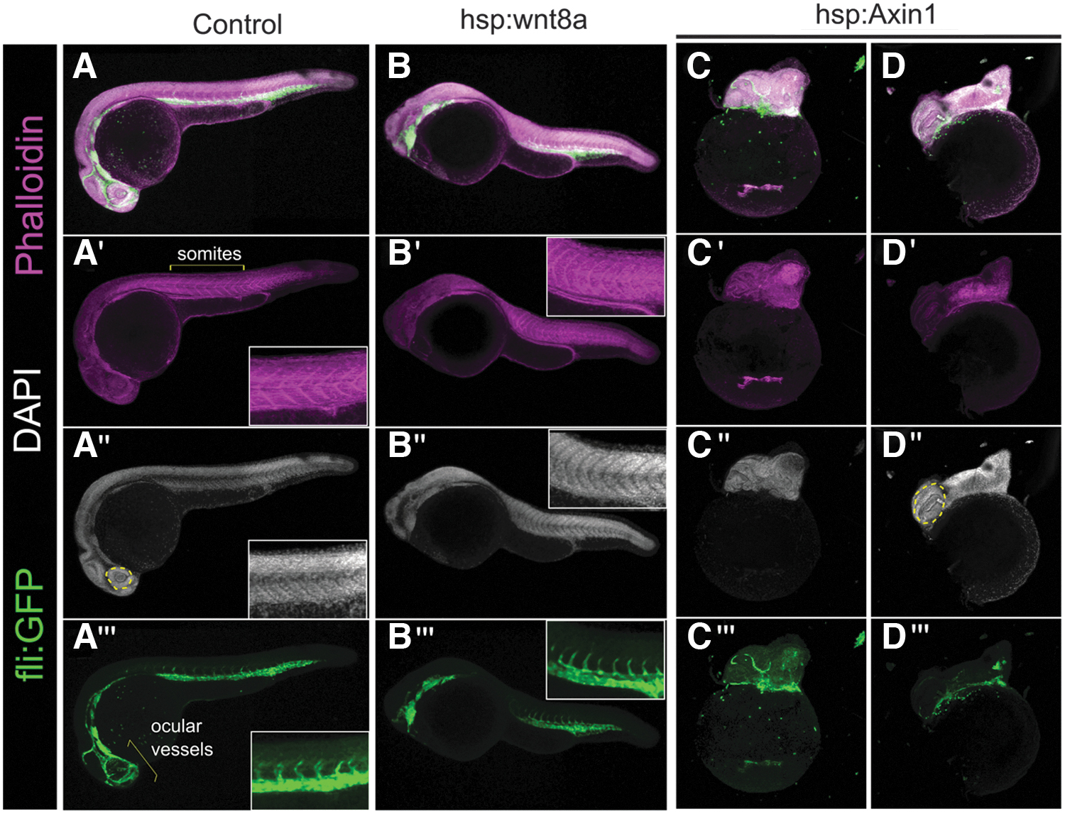
Fig. 5 Student-generated confocal images of 24 hpf embryos following Wnt pathway manipulation. Top row, merged images of DAPI (gray), Phalloidin (magenta) and fli:GFP (green), A′–D‴ individual channels of control (A), HS hsp:wnt8a (B), and HS hsp:Axin1 (C, D). HS was performed for 40 min at 37 degrees starting at sphere stage (B, D) or 1000–2000 cell (3 hpf) stage (C). Wnt overexpression reduces eye development, and accompanying ocular vessels are absent (A‴, bracket vs. B‴). Inset highlights trunk tissue patterning based on phalloidin (A′, B′) nuclear staining (A″, B″) and intersegmental vessel outgrowth (A‴, B‴). Wnt inhibition causes increased size of eye (yellow dashed outline, A″ vs. D″) and disrupts posterior axis outgrowth (C, D). Figure “A” is a stitched image of two partially overlapping images (see Materials and Methods section for details).