
IMAGE
Fig. 4
- ID
- ZDB-IMAGE-240524-63
- Publication
- Fiegl et al., 2024 - Laboratory Course Using Zebrafish to Uncover Changing Roles of Wnt Signaling in Early Vertebrate Development
- All Figures
- Figures for Fiegl et al., 2024
Image
|
Figure Caption
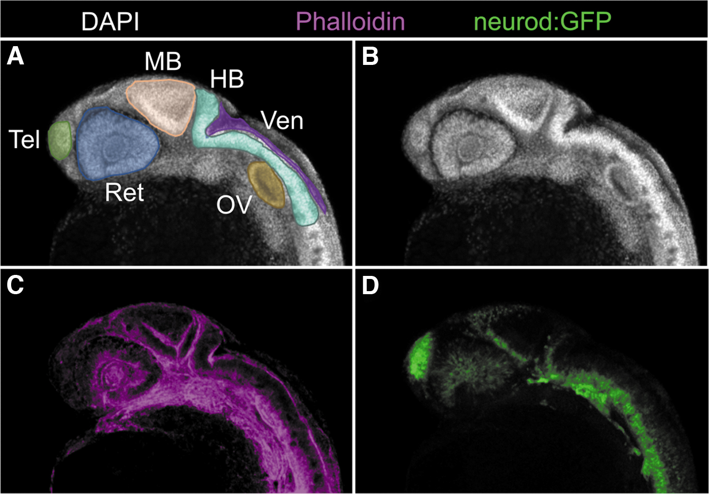
Fig. 4 CNS morphology revealed by DAPI (gray) and phalloidin (magenta) staining, and neurod:GFP (green) at 24 hpf. (A, B) Nuclear staining highlights anterior structures. Phalloidin staining further outlines structures (C), complementing information provided by transgenes such as neurod:GFP (green, D). CNS; DAPI; HB, hindbrain; MB, midbrain; OV, otic vesicle; Ret, retina; Tel, telencephalon; Ven, hindbrain ventricle.
Acknowledgments
This image is the copyrighted work of the attributed author or publisher, and
ZFIN has permission only to display this image to its users.
Additional permissions should be obtained from the applicable author or publisher of the image.
Full text @ Zebrafish