
IMAGE
Fig. 3
- ID
- ZDB-IMAGE-240524-62
- Publication
- Fiegl et al., 2024 - Laboratory Course Using Zebrafish to Uncover Changing Roles of Wnt Signaling in Early Vertebrate Development
- All Figures
- Figures for Fiegl et al., 2024
Image
|
Figure Caption
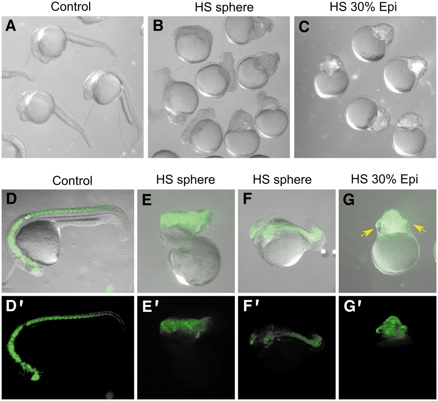
Fig. 3 Reduced posterior extension in HS hsp:Axin1 transgenic embryos. (A, B) Brightfield images at 24 hpf of control (A) and hsp:Axin1 embryos that received a HS at sphere stage (B) and at 30% epiboly (C). (D–G) neurod:GFP with brightfield overlay of control embryos (D) or hsp:Axin1 embryos treated with HS as indicated (E–G). (D′–G′) Neurod: GFP channel shown alone. Eye formation was evident in severely truncated embryos (G, yellow arrows).
Acknowledgments
This image is the copyrighted work of the attributed author or publisher, and
ZFIN has permission only to display this image to its users.
Additional permissions should be obtained from the applicable author or publisher of the image.
Full text @ Zebrafish