
|
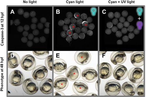
Fig. 6 Reversible Embryo Growth Arrest with E-1 and Two Light Frequencies. Embryos were treated with 0.2 μM of E-1 at 4 hpf and immediately exposed to cyan light (490 nm, B–C, E–F) for 60 minutes to produce mostly Z-1 or left non-irradiated as control (A, D). At 5.5 hpf, part of the embryos was exposed to 365 nm irradiation for 12 minutes to convert most of the Z-1 into its less toxic E-isomer (C, F). Then, all embryos were rinsed with E3 medium at 6.5 hpf, to remove plinabulin from the environment. Afterwards, one group of embryos was incubated until 12 hpf and subsequently fixed and their apoptotic cells were stained (immunohistochemistry using anti-activated caspase-3 antibody; white spots; A–C). The other group was incubated until 48 hpf to analyze their phenotype for long-term development after treatment (D–F). The group treated without inactivation (exposure to the more toxic Z-1 isomer for 2.5 hours) displays a pronounced number of apoptotic cells at 12 hpf and predominantly malformed embryos with improperly developed heads and tails at 48 hpf (B, E; red asterisks). Embryos inactivated using UV light (365 nm) at 5.5 hpf (1.5 hours exposure to Z-1) exhibit a low number of apoptotic cells at 12 hpf and correctly developed heads, tails, and regular internal structure at 48 hpf (C, F).