
|
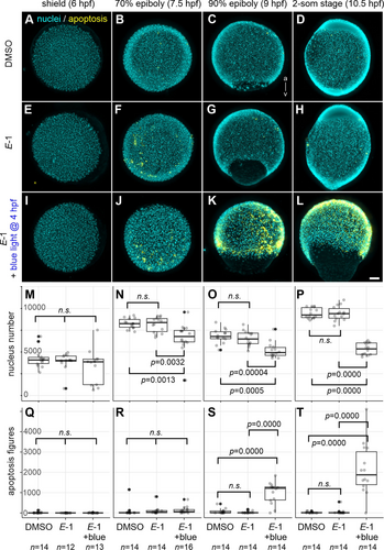
Fig. 5 Cyan Light-Induced Mitotic Arrest Followed by Apoptosis in E-1 Treated Embryos. Zebrafish embryos at 4 hpf were transferred to E3 medium containing DMSO (A–D) or E-1 (E–L). A subset of E-1-treated embryos underwent cyan light (490 nm) exposure for 1 hour (I–L). All embryo groups were subsequently grown in darkness until either 6 hpf (shield stage; A, E, I), 7.5 hpf (70 % epiboly; B, F, J), 9 hpf (90 % epiboly; C, G, K) or 10.5 hpf (2-som stage; D, H, L) and fixed for the analysis of nuclei number (DAPI staining; cyan) and apoptotic figures (immunohistochemistry using anti-activated caspase-3 antibody; yellow). Embryos were oriented either for the animal pole view (A–B, E–F, I–J) or the lateral view with the animal pole up and dorsal right (C–D, G–H, K–L). (M–P) Number of nuclei per individual embryo. Note that the absolute values from different stages are not directly comparable because of the fact that only one-third of the embryo was captured in 3D volumetric imaging. (Q–T) Number of apoptotic figures per individual embryo. n.s.: not significant; Scale bar: 100 μm; n >12.