
|
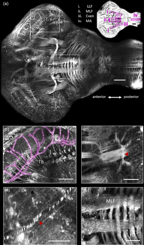
Fig. 3 Identification of major fiber bundles and vasculature with third harmonic generation microscopy in the adult brain of Danionella dracula. (a) Maximum projection of all the frames that contain the medial longitudinal fasciculus (MLF). Anterior-posterior axis is indicated. Color-inverted image is presented in top right corner with major fiber bundles outlined in magenta (see adjacent legend). Scale bar indicates 100 μm for both images. LLF: lateral longitudinal fasciculus; Cven: ventral commissure; MA: Mauthner axon. (b) Region of optic tectum (TeO) outlined in (a). This image contains blood vessels (false colored with magenta) as well as individual axon fascicles that form the tectobulbar fiber bundles (TB). (c) Example of fast flowing red blood cells (RBCs) in blood vessel (red arrowhead). (d) Example of slow flowing RBCs in blood vessel (red arrowhead). This image is an average of several images, each containing a single red blood cell, leading to each cell being distinctly visible. (e) Higher magnification view of MLF and Cven in hindbrain outlined in (a). Scale bars indicate 50 μm for (b–e). Images collected with a pixel size of 0.53 μm (a, b, e) and 0.26 μm (c, d).