
|
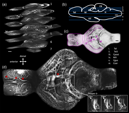
Fig. 2 Representative images obtained by third harmonic generation microscopy through the entire depth of the brain of an adult Danionella dracula. Whole-brain images were obtained by stitching together stacks from five field of views (see Section 2). (a) Three-dimensional distribution of structures at seven horizontal levels within the brain. Compass indicates brain direction for (a–d). Images were collected with a pixel size of 0.53 μm. Scale bar indicates 100 μm in the xy plane. Numbers on each plane represent the depth inside the brain as depicted in panel (b). (b) Horizontal planes in part (a) are labeled at their approximate depth on sagittal line drawing of the brain with blue lines. Labels on line drawing indicate telencephalon (Tel), midbrain (M), cerebellum (CB), hindbrain (H), and hypothalamus (HY). Scale bar indicates 100 μm. (c) Identification of prominent myelinated fiber bundles of image in (d). Image is the same as panel (d) and presented in inverted color scale for clarity. Tel: telencephalon, TeO: Optic tectum, Cpost: posterior commissure, MV: midbrain ventricle, Ccer: Cerebellar commissure, Cgus: gustatory commissure. Scale bar indicates 100 μm. (d) Maximum projection of all the frames that contain Cpost. Red arrowheads indicate blood vessels. Panel on the bottom left shows progressively deeper (left to right) individual frames from the stack going through the Ccer and Cgus. Scale bar indicates 100 μm.