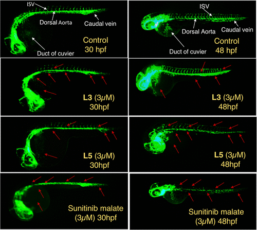
Fig. 12 In vivo antiangiogenic effects of L3 (3 μM) and L5 (3 μM) in Tg(Fli1:gfp) Zebrafish through in vivo experiments conducted at various time intervals. The compound addition was done at 14hpf and incubation continued for 12 h. The GFP-expressing regions of interest, including the duct of Cuvier, dorsal aorta, ISVs, and caudal vein, are indicated by white arrows in the control group treated with 0.2% DMSO. Sunitinib malate (3 μM) was taken as the positive control. Conversely, the affected areas, reflected by the loss of the GFP-expressing vasculature due to the antiangiogenic effect in the treated samples, are identified by red arrows. The data show that between L3 and L5, the most effective one is L5 at just a 3 μM concentration, showing a strong antiangiogenic effect even at 48 hpf when the zebrafish recovered partly from the effect of L3. The commercial drug sunitinib malate used as standard shows the strongest effect.
Image
Figure Caption
Acknowledgments
This image is the copyrighted work of the attributed author or publisher, and
ZFIN has permission only to display this image to its users.
Additional permissions should be obtained from the applicable author or publisher of the image.
Full text @ J. Med. Chem.

