
Image
|
Figure Caption
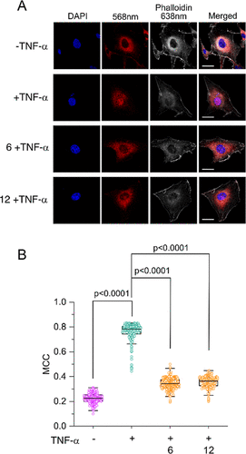
Fig. 8 (A) Representative confocal images of HMEC-1 cells showing TNF-α-mediated NF-κB nuclear localization after treatment with the indicated complex (10 μM) for 2 h followed by activation with TNF-α for 1 h, as indicated in each panel. F-actin and the nucleus are stained with phalloidin (gray) and DAPI (blue), respectively. Scale bar, 30 μm. (B) Quantification of colocalization between NF-κB and DAPI by MCC at the indicated experimental condition. (Cell number = 150–180) in the cytoplasm of HMEC-1 cells suggesting that the complexes inhibit the nuclear translocation of NF-κB.
Acknowledgments
This image is the copyrighted work of the attributed author or publisher, and
ZFIN has permission only to display this image to its users.
Additional permissions should be obtained from the applicable author or publisher of the image.
Full text @ J. Med. Chem.