
|
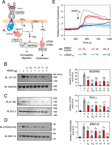
Fig. 5 (A) Schematic representation of VEGFR2 signaling in angiogenesis. (64,65) Signaling modules probed are highlighted (KEGG pathway database). (B–D) In the left panel is the representative Western blot showing the level of VEGFR2(B), PLC-γ (C), and ERK1/2 (D) phosphorylation after treating the HMEC-1 cells with the indicated compounds (10 μM) for 6 h followed by activation with VEGF165 (30 ng/mL). In the right panel, the bar plots represent the densitometric quantification of Y1175(B), Y783(C), or T202/Y204(D) phosphorylation level after compound treatment. The error bar is the standard deviation from three independent experiments. (E) Ca2+ flux is determined from the plot of change in fluorescence of Fluo-4 as a function of time in HMEC-1 cells treated with the indicated compounds (10 μM) for 3 h followed by activation with VEGF165 (30 ng/mL).