IMAGE

Fig. 4

ID
ZDB-IMAGE-240515-37
Source
Figures for Hao et al., 2024
Image
Figure Caption

Fig. 4

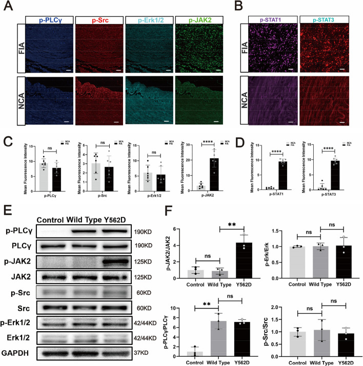
The JAK2-STAT pathway serves as the major downstream signaling pathway of the mutated PDGFB. mIF demonstrate the important downstream signaling pathway markers of PDGFRβ (p-JAK, p-Src, p-Erk1/2 and p-PLCγ) in FIA sections and NCA sections (A). Scale bar, 100 μm. Statistical analysis is performed to compare the mean fluorescence intensity of different signal markers between NCAs (n = 4) and FIAs (n = 5) (C). The expression of p-STAT1 and p-STAT3, which are downstream of p-JAK2, is detected by mIF in FIAs and NCAs. Scale bar, 50 μm (B). Statistical analysis of mean fluorescence intensity of these marks between NCAs (n = 4) and FIAs (n = 5) (D). Western blotting displays the expression of the important downstream signal pathway markers of PDGFRβ (p-JAK2, p-Src, p-Erk1/2 and p-PLCγ) in the HBVSMCs underwent different treatment in vitro (E). Relative density (normalized to Control) of immunoblot bands from (E) corresponding to pPLCγ, p-JAK2, p-Src, and p-ERK1/2 (n = 3) (F). 'n' represented the number of samples. Three random fields were selected for statistical analysis in each sample, and the average value represented the detection value of this marker in this sample. Tukey's multiple comparisons test is conducted to evaluate the statistical differences. ns, no significant; ***padj < 0.001, ****padj < 0.0001

Acknowledgments
This image is the copyrighted work of the attributed author or publisher, and ZFIN has permission only to display this image to its users. Additional permissions should be obtained from the applicable author or publisher of the image. Full text @ J. Biomed. Sci.