
|
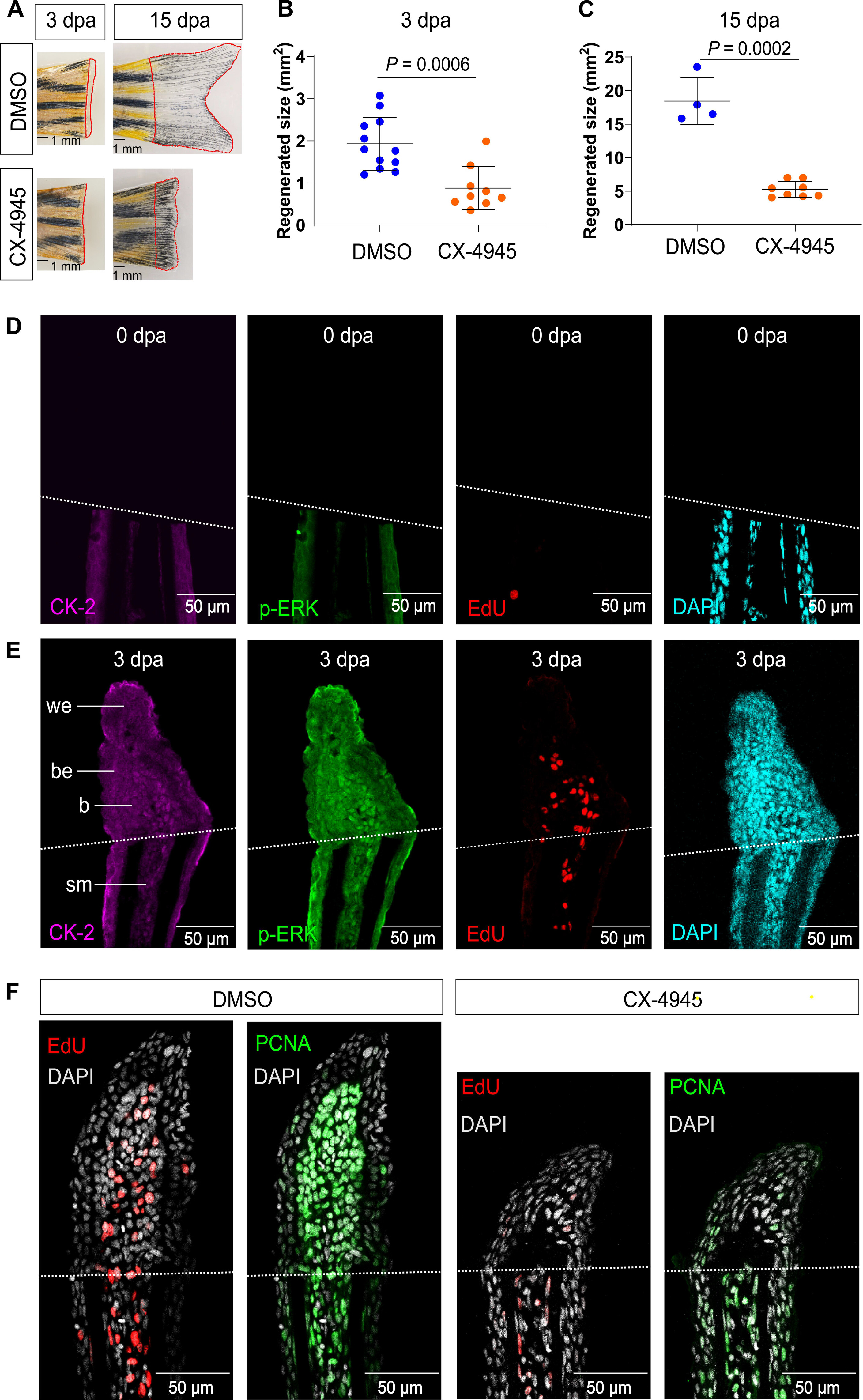
Fig. 6 CK-2 drives blastema formation in zebrafish by promoting cell proliferation. (A) Regenerating fins at 3 and 15 dpa after dimethyl sulfoxide (DMSO) or CX-4945 (20 μM) treatment. The red frames indicate the regenerated area. Scale bars, 1 mm. (B and C) Quantification of fin regenerating tissue size from (A). Student’s t test. (D and E) Sections of regenerating fins stained with CK-2, p-ERK, and EdU at 0 and 3 dpa. The white dashed line indicates amputation plane. we, wound epidermis; be, basal epidermal layer; b, blastema; sm, stump mesenchyme. Scale bars, 50 μm. (F) Inhibition of CK-2 blocks the blastema formation by down-regulating the cell proliferation. EdU staining (red) and PCNA antibody staining (green) were used to examine cell proliferation. Nuclei were stained using DAPI (white). The white dashed line indicates amputation plane. Scale bars, 50 μm.