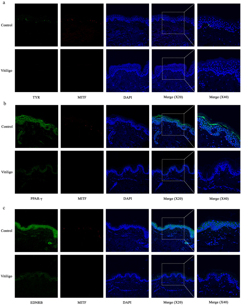
IMAGE

Fig. 3

ID
ZDB-IMAGE-240508-39
Source
Figures for Zhao et al., 2023
Image
Figure Caption

Fig. 3

Melanocyte deficiency in vitiligo lesions is accompanied by impaired expression of PPAR-γ and EDNRB. a Representative images of the melanocytes in healthy skin samples (n = 5) and vitiligo lesions (n = 5) detected by immunofluorescence. Melanocytes were stained with antibodies to TYR (green) and MITF (red). Nuclei were counterstained with DAPI (blue). b Representative images of the expression of PPAR-γ (green) in healthy skin (n = 5) and vitiligo lesions (n = 5) detected using immunofluorescence. Melanocytes were stained with antibodies to MITF (red). Nuclei were counterstained with DAPI (blue). c Representative images of the expression of EDNRB (green) in healthy skin (n = 5) and vitiligo lesions (n = 5) detected using immunofluorescence. Melanocytes were stained with antibodies to MITF (red). Nuclei were counterstained with DAPI (blue).

Acknowledgments
This image is the copyrighted work of the attributed author or publisher, and ZFIN has permission only to display this image to its users. Additional permissions should be obtained from the applicable author or publisher of the image. Full text @ Inflammation