
|
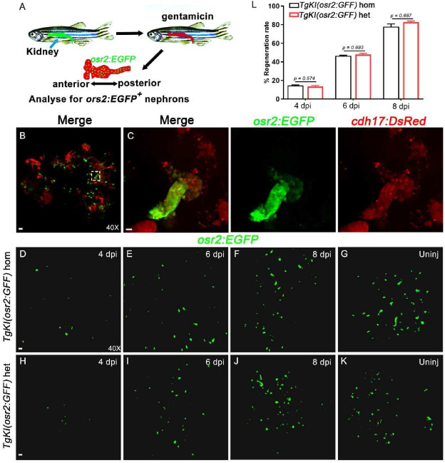
Fig. 6 EGFP + cells regeneration in Tg(osr2:EGFP) adult zebrafish after kidney injury. A: Schematic diagram of the AKI model constructed by intraperitoneal injection of gentamicin in adult zebrafish. B: Confocal images of adult zebrafish PCT cells that are co-labeled by osr2:EGFP and cdh17:DsRed are shown (n = 6). Scale bar in A, 100 μm. C: Higher-magnification image of the boxed area showed in B. Scale bar, 200 μm. D-G: Use of osr2:EGFP background to show of PCTs in TgKI(osr2:GFF) homozygotes at the indicated time point after injured (n = 6). Scale bar, 100 μm. H–K: Use of osr2:EGFP background to show of PCTs in TgKI(osr2:GFF) heterozygotes at the indicated time point after injured (n = 6). Scale bar, 100 μm. L: After injured, the renal regeneration rate of TgKI(osr2:GFF) homozygotes and TgKI(osr2:GFF) heterozygotes were counted using ImageJ (n = 5 biological replications per group). hom, homozygotes; het, heterozygotes. Scale bar in A to J, 500 μm.