IMAGE

Fig. 4

ID
ZDB-IMAGE-240505-4
Source
Figures for El Omar et al., 2024
Image
Figure Caption

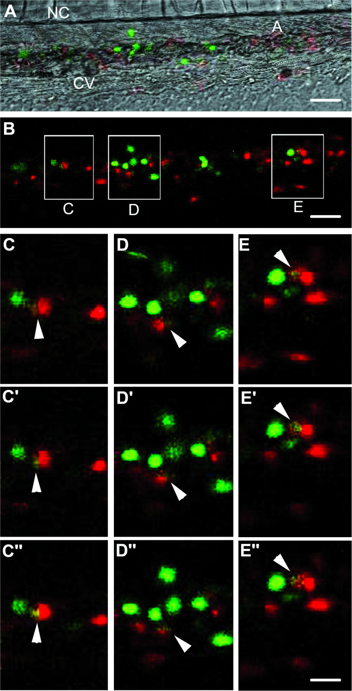
Fig. 4 CD34-GFP cells phagocytosis by zebrafish embryonic macrophages.

A, B Z-stack confocal imaging of mCherry-expressing macrophage behavior in a zebrafish embryo 2 h post transplantation (h.p.T.) of CD34-GFP, reveals the accumulation of macrophages around green cells and their phagocytosis (inset C, D and E). A Bright field imaging of caudal hematopoietic tissue in the tail region at 30 h.p.f., merged with confocal fluorescent imaging of CD34-GFP cells and mCherry-expressing macrophages (B). CE” 3 successive z-stacks confocal planes apart 0.8 μm of phagocytosis of CD34-GFP cells by mCherry-expressing macrophage (arrows). See also Video 6. A aorta, CV caudal vein, NC notochord. Scale bars: A, B: 25 µm; CE”:10 µm.

Acknowledgments
This image is the copyrighted work of the attributed author or publisher, and ZFIN has permission only to display this image to its users. Additional permissions should be obtained from the applicable author or publisher of the image. Full text @ Cell Death Dis.