IMAGE

Fig. 2

ID
ZDB-IMAGE-240505-2
Source
Figures for El Omar et al., 2024
Image
Figure Caption

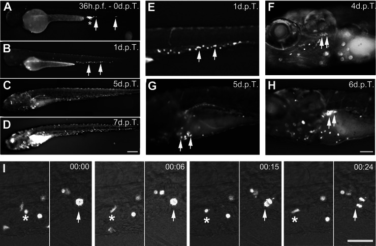
Fig. 2 Transplantation of human JK-GFP cells in zebrafish embryos, colonization of hematopoietic organs and proliferation.

A Transplantation of JK-GFP cells in the caudal vein of a wildtype zebrafish embryo at 36 h.p.f. BD Colonization of zebrafish hematopoietic tissues followed from 1 to 7 d.p.T. Colonization of the CHT (B, E, arrows), thymus (F, arrows), fetal liver (G, arrows), kidney (H, arrows). See Video 1. I Live imaging of JK-GFP cells circulation (asterisk) and proliferation (arrow) in the CHT (Time code 00:15 and 00:24). See Video 2. CHT: Caudal Hematopoietic Tissue. Scale bars: (AD): 250 µm; (EH):100 µm, (I): 25 µm. Time code in minutes.

Acknowledgments
This image is the copyrighted work of the attributed author or publisher, and ZFIN has permission only to display this image to its users. Additional permissions should be obtained from the applicable author or publisher of the image. Full text @ Cell Death Dis.