IMAGE

Fig. 1

ID
ZDB-IMAGE-240505-1
Source
Figures for El Omar et al., 2024
Image
Figure Caption

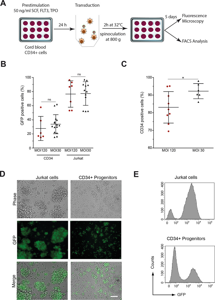
Fig. 1 Transgene transfer and GFP expression after transduction of human CD34+ and Jurkat cells.

A Human CD34+ blood cells from umbilical cord blood and Jurkat cells were transduced by spin inoculation with lentiviral vectors encoding a nuclear variant of the eGFP at multiplicity of infection (MOI) of 30 and 120 and cultured in vitro. After 5 days, cells were analyzed for GFP expression by both flow cytometry and fluorescence microscopy. B Estimates of the efficiency of transduction based on the percentage of GFP+ cells. C Percentage of cells expressing CD34 after 5 days of culture. D, E A representative experiment showing GFP expression after transduction of human CD34+ or Jurkat cells at the MOI of 30. In (E), results are represented as histograms of GFP fluorescence intensity (x-axis, log scale) versus cell number (y-axis, linear).

Acknowledgments
This image is the copyrighted work of the attributed author or publisher, and ZFIN has permission only to display this image to its users. Additional permissions should be obtained from the applicable author or publisher of the image. Full text @ Cell Death Dis.