IMAGE

Fig. 6

ID
ZDB-IMAGE-240429-51
Source
Figures for Liu et al., 2024
Image
Figure Caption

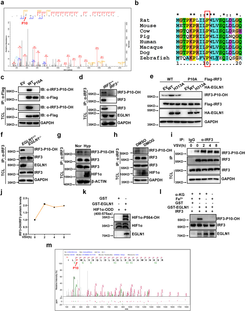
Fig. 6 EGLN1 hydroxylates IRF3 at proline 10.

a The hydroxylated residue in IRF3 was identified by mass spectrometry analysis. b Sequence alignment of partial IRF3 (1–20 amino acids). c HEK293T were transfected with the Flag empty vector (EV), Flag-IRF3 or Flag-IRF3-P10A, followed by immunoprecipitation and immunoblotting with the indicated antibodies. d Cell lysates from IRF3+/+ and IRF3−/− H1299 were extracted, followed by immunoprecipitation and immunoblotting with the indicated antibodies. eEGLN1−/− H1299 were transfected with Flag-IRF3-WT or Flag-IRF3-P10A, together with the HA empty vector (EV), HA-EGLN1-WT (WT), HA-EGLN1-H313A (H313A), followed by immunoprecipitation and immunoblotting with the indicated antibodies. f Cell lysates from EGLN1+/+ and EGLN1−/− H1299 were extracted, followed by immunoprecipitation and immunoblotting with the indicated antibodies. g Cell lysates from THP-1 under normoxia (21% O2) or hypoxia (1% O2) for 12 h were extracted, followed by immunoprecipitation and immunoblotting with the indicated antibodies. h Cell lysates from THP-1 treated with DMSO (vehicle control) or DMOG (1 mM) for 6 h were extracted, followed by immunoprecipitation and immunoblotting with the indicated antibodies. i Cell lysates from H1299 infected with VSV virus for the indicated time were extracted, followed by immunoprecipitation and immunoblotting with the indicated antibodies. j Quantitation of the intensity of hydroxylated IRF3/total IRF3 protein in (i). k Bacterially expressed HIF1α (400-575aa) (GST tag has been removed using thrombin) incubated with bacterially expressed GST or bacterially expressed GST-EGLN1 for hydroxylation reaction and then detected by immunoblotting with anti-HIF1α-P564-OH antibody. l In vitro hydroxylation assay to detect bacterially expressed IRF3 hydroxylated by bacterially expressed GST-EGLN1. m Bacterially expressed IRF3 (GST tag has been removed using thrombin) was incubated with bacterially expressed GST-EGLN1 for hydroxylation reaction and then identified by mass spectrometry analysis. Hydroxylated IRF3-P10 is indicated by the red arrow, and the percentage of IRF3 hydroxylation is 10.34%. EV empty vector, IP immunoprecipitation, TCL total cell lysates. Data in (ci, k, l) are representative from three independent experiments. See also Supplementary Fig. 11. Source data are provided as a Source data file.

Acknowledgments
This image is the copyrighted work of the attributed author or publisher, and ZFIN has permission only to display this image to its users. Additional permissions should be obtained from the applicable author or publisher of the image. Full text @ Nat. Commun.