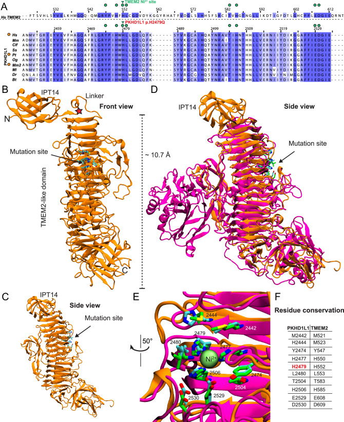
IMAGE

Fig. 4

ID
ZDB-IMAGE-240426-81
Source
Figures for Redfield et al., 2024
Image
Figure Caption

Fig. 4

PKHD1L1 structural modeling of the protein fragment containing the p.(His2479Gln) variant. Based on AlphaFold2 predictions, this fragment of PKHD1L1 shares a common fold with the TMEM2 protein within the region carrying the p.(His2479Gln) variant. a Protein sequence alignment of a protein segment of Hs TMEM2 against ten different PKHD1L1 orthologs (see Supplementary Table S1 for details of the selected species, Supplementary Fig. S4 for sequence alignment of this specific fragment, and Supplementary Fig. S3 for full PKHD1L1 sequence alignment). Residue numbering for TMEM2 as in PDB: 8C6I (Niu et al. 2023), while residue numbering for Hs PKHD1L1 as in NCBI accession code NP_803875.2 with the signal peptide included (Supplementary Table S1, 26 residues are suggested according to protein sequence alignment, see Methods). Green triangles point to the location of the Hs p.(His2479Gln) variant, orange circles (left) indicate 100% amino acid sequence identity for this PKHD1L1 fragment between the Hs, Pt, and Mm2 species (See supplementary Table S1 for details about the selected orthologs). Green circles represent depicted residues in panels b–e. The alignment was color-coded for sequence similarity (35% threshold) using Jalview. White-colored residues show the lowest similarity and dark blue report the highest (see Methods). PKHD1L1 orthologs were chosen based on sequence availability and taxonomical diversity. b The simulated protein structure covering the protein fragment highlighted by purple arrow-headed line in Fig. 2b. Front view of the structure showing IPT14 linked to the PKHD1L1 TMEM2-like domain. The red star points to the linker connection. Residues at the mutation site are depicted as cyan sticks. c Side view from panel a showing a clear view of the stacked β-strand motifs. d Superposed structural protein alignment between WT Hs PKHD1L1 TMEM2-like domain model (orange) with the X-ray crystal structure of Hs TMEM2 protein (PDB: 8C6I, magenta). Residues at the native TMEM2-histidine-finger site are depicted as green sticks. e Higher magnification image of the potential conserved histidine-finger site between PKHD1L1 (orange) and TMEM2 (magenta) protein fragments and the Ni2+ ion shown as lime sphere. f Displayed residues between both proteins surrounding the Ni2+-ion site highlighted in panel a in green circles

Acknowledgments
This image is the copyrighted work of the attributed author or publisher, and ZFIN has permission only to display this image to its users. Additional permissions should be obtained from the applicable author or publisher of the image. Full text @ Hum. Genet.