
|
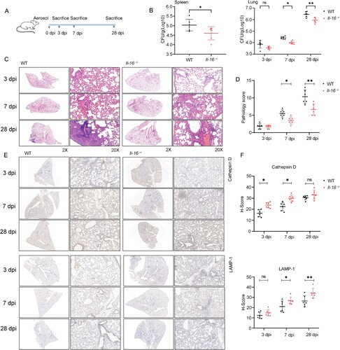
Fig. 7 Interleukin 16 deficiency reduced the host susceptibility to H37Rv infection. A Schematic diagram of infection experiments. C57BL/6 or Il-16-/- mice were infected with ∼200 CFU of H37Rv using a Glas-Col inhalation exposure system (n = 6 to 8). B At 3-, 7- and 28-days post-infection, the mice were sacrificed, and bacterial counts in the lungs and spleens were determined on Middlebrook 7H10 agar. Mtb colonies were incubated at 37 °C and counted after 21 days (n = 6). C For histopathology analysis, half of each lung was fixed in a 4% neutral-buffered paraformaldehyde solution for 24 hours. Lung tissue was then embedded in paraffin. A series of sections with a thickness of 4-7 μm were then cut and stained with hematoxylin and eosin by standard methods. D Histopathology analysis was evaluated by pathologists in a blinded manner (n = 6). E LAMP-1 and Cathepsin D expression were analyzed by immunohistochemistry. F H-Score analysis was determined according to E. The values are the means ± SEM, 6 to 8 mice per group as indicated. *p<0.05 compared to media, Student's t-test.