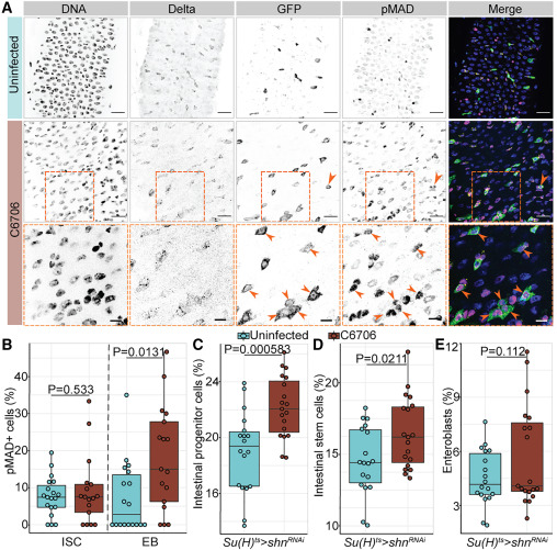
Fig. 5 Vc-responsive BMP activation in EBs arrests intestinal stem cell growth non-cell autonomously (A) Posterior midguts of Su(H)ts/+ adult flies uninfected or infected with C6706 for 36 h. Hoechst labels DNA (blue); Delta marks ISCs (yellow); GFP labels Su(H)+ EBs (green); pMAD labels cells with BMP activation (magenta). Arrows and dashed box: pMAD+ GFP+ EBs. Scale bars in dashed boxes: 5 μm. Scale bars in images: 15 μm. (B) Proportion of intestinal stem cells or EBs that are pMAD+ in Su(H)ts/+ intestines. (C–E) Proportions of all cells that are (C) progenitors, (D) ISCs, or (E) EBs in the intestines with EB-specific BMP inactivation (Su(H)ts>shnRNAi). Each dot represents a measurement from a single fly gut. p values are calculated using unpaired Student’s t tests.
                
                    
                        Image


                        Figure Caption


                        Acknowledgments


This image is the copyrighted work of the attributed author or publisher, and
ZFIN has permission only to display this image to its users.
Additional permissions should be obtained from the applicable author or publisher of the image.
      Full text @ Cell Rep.


        