
|
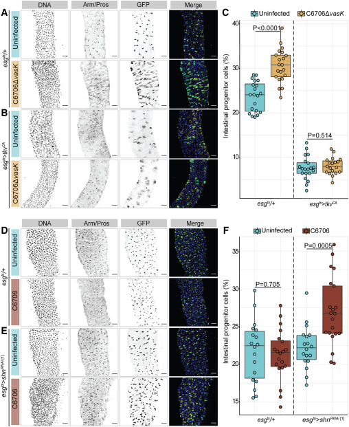
Fig. 4 BMP regulates epithelial repair after Vc infection (A and B) Posterior midguts from (A) WT (esgts/+) or (B) progenitor-specific BMP activation (esgts>tkvCA) adult flies uninfected or infected with C6706ΔvasK for 36 h. DNA labeled with Hoechst (blue); GFP marks progenitors (green); and Arm/Pros label cell borders and enteroendocrine cells, respectively (yellow). Scale bar: 25 μm. (C) Proportion of intestinal epithelial cells that are GFP+ progenitors in esgts/+ and esgts>tkvCA flies. (D and E) Images of (D) WT (esgts/+) or (E) progenitor-specific BMP inhibition (esgts>shnRNAi) adult flies uninfected or infected with C6706 for 36 h. DNA labeled with Hoechst (blue); GFP marks progenitors (green); and Arm/Pros label cell borders and enteroendocrine cells, respectively (yellow). Scale bar: 25 μm. (F) Proportion of cells that are GFP+ progenitors in esgts/+ and esgts>shnRNAi flies. Each dot represents a measurement from a single fly gut. p values are calculated using unpaired Student’s t tests.