
|
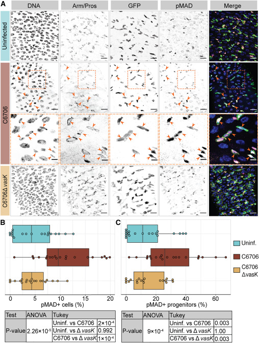
Fig. 1 Vc activates BMP in intestinal progenitor cells in a T6SS-dependent manner (A) Posterior midguts of esgts/+ adult fly uninfected or infected with C6706 or C6706ΔvasK for 24 h. Hoechst labels DNA (blue); Armadillo and Prospero (Arm/Pros) label cell borders and enteroendocrine cells, respectively (yellow); GFP labels progenitors (green); and pMAD labels cells with BMP activation (magenta). Arrowheads: pMAD+ GFP+ progenitors. The area within the dashed box of row two is presented at a higher magnification in row three to better illustrate pMAD in progenitors. Scale bars in row three: 5 μm. All other scale bars: 15 μm. (B and C) Percentage of (B) all cells or (C) progenitors that are pMAD+. Each dot represents a measurement from a single fly gut. p values are calculated using the significance tests indicated in the tables