
Fig. 5
- ID
- ZDB-IMAGE-240417-18
- Publication
- Lin et al., 2023 - Extracellular Matrix Disorganization Caused by ADAMTS16 Deficiency Leads to Bicuspid Aortic Valve With Raphe Formation
- All Figures
- Figures for Lin et al., 2023
|
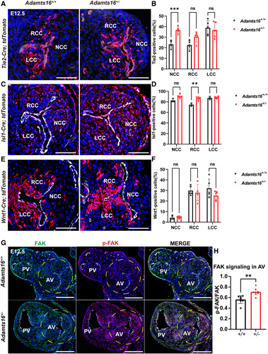
Fig. 5 The effects of Adamts16 deficiency on the activities of each cell lineage during the development of the aortic valve. A through F, Fate mapping of the endothelial ( A), neural crest ( C), and second heart field ( E) lineage cells was performed respectively in the aortic valves of both Adamts16+/+ and Adamts16+/- embryos at E12.5. Transversal sections were immunoassayed with PECAM1 (in white) and DAPI (in blue). Scale bars=100 µm. Quantification of endothelial-derived cells demonstrates the increased cell numbers in the noncoronary cusp in Adamts16+/- (n=5) compared with Adamts16+/+ littermates (n=5; **P<0.01; B). Quantification of second heart field–derived cells demonstrates increased cell numbers in the noncoronary and right coronary cusps in Adamts16+/- mice (n=5) compared with Adamts16+/+ littermates (n=3). **P<0.01; ****P<0.0001 ( D). Quantification of neural crest–derived cells demonstrates that there was no significant difference in cell number in 3 individual cups of Adamts16+/- (n=5) compared with Adamts16+/+ littermates (n=5) ( F). In B, D, and F, Statistical analysis was performed using 2-way ANOVA followed by the Šidák multiple comparisons test. G, Immunostaining of FAK and phospho-FAK (Tyr397) in aortic valves of Adamts16+/+ and Adamts16+/- embryos at E12.5. Scale bars=100 µm. H, Quantification of phospho-FAK (Tyr397) staining (n=8 for each genotype). P values were calculated using the Student t test. ADAMTS indicates a disintegrin and metalloproteinase with thrombospondin motifs; AV, aortic valve; LCC, left coronary cusp; NCC, noncoronary cusp; PV, pulmonary valve; and RCC, right coronary cusp.