
|
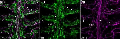
Fig. 3 Confocal imaging of immunohistochemical co-localization of tyrosine hydroxylase (TH) with a zebrafish-specific neuronal marker. (a) Labeling with anti-TH (green) co-localized with nerve fibers labeled with zn-12 (magenta). (b, c) TH and zn-12 labeling shown separately. Both nerve bundles of the central filament (F) and all zn-12-positive nerve fibers (arrowheads) of the filaments and lamellae (L) were also TH positive. Some labeling by anti-TH did not co-localize with zn-12. An intrabranchial chain neuron (ChN) is indicated that was labeled with both markers. Scale bar in panel (a) = 10 μm and applies to all panels.