
|
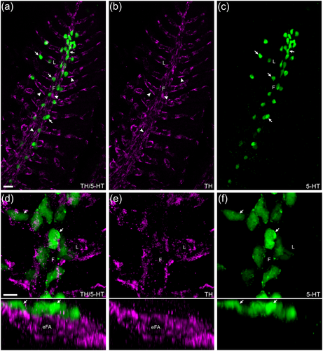
Fig. 2 Confocal imaging of immunohistochemical localization of tyrosine hydroxylase (TH)-positive nerve fibers associated with serotonergic neuroepithelial cells (NECs). (a) NECs (arrows) labeled with anti-serotonin (5-HT, green) were closely associated with a plexus of nerve fibers (arrowheads) labeled by anti-TH (magenta) in the filaments (F) and lamellae (L). (b, c) TH and 5-HT labeling shown separately. (d–f) Images of gill filaments and lamellae are shown at higher magnification (upper panels) and tilted back 90° (lower panels). In the latter, the transverse optical section revealed the close association between NECs and nerve fibers, and that the nerve fibers surrounded the efferent filament artery (eFA). Scale bar in panel (a) = 20 μm and applies to panels (b) and (c). Scale bar in panel (d) = 20 μm and applies to panels (e) and (f).