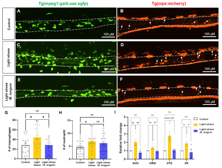
Probiotics reduced inflammatory cell recruitment by constant light exposure. (A,C,E) In vivo intestinal images of 10 dpf Tg (mpeg1:gal4vp16;uas:egfp) show macrophage infiltration. Scale bar: 100 μM. (B,D,F) In vivo intestinal images of 10 dpf Tg (mpx:mcherry) show neutrophil infiltration. Arrows indicate neutrophils in the intestine. Scale bar: 100 μM. (A,B) Control group. (C,D) Stress-induced group by constant light exposure. (E,F) B. longum-feeding stress-induced group. (G) Comparison of the number (#) of intestine-infiltrated macrophages among all groups: n = 16 for control group; n = 21 for light-stress group; n = 13 for light-stress/B. longum group. (H) Comparison of the number (#) of intestine-infiltrated neutrophils among all groups: n = 18 for control group; n = 21 for light-stress group; n = 21 for light-stress/B. longum group. (I) Relative fold changes of inflammatory genes, including tnfα, nfkb, il1b, and il6 (n = 4). Each replicate had 20 zebrafish embryos. ns, not significant; * p < 0.05; ** p < 0.01; *** p < 0.001; **** p < 0.0001.
Acknowledgments
This image is the copyrighted work of the attributed author or publisher, and
ZFIN has permission only to display this image to its users.
Additional permissions should be obtained from the applicable author or publisher of the image.
Full text @ Int. J. Mol. Sci.
Your Input Welcome
Thank you for submitting comments. Your input has been emailed to ZFIN curators who may contact you if
additional information is required.
Oops. Something went wrong. Please try again later.